2VDT

Crystallographic structure of Levansucrase from Bacillus subtilis mutant S164A


Experimental Data Snapshot

  • Method: X-RAY DIFFRACTION
  • Resolution: 3.20 Å
  • R-Value Free: 0.297 
  • R-Value Work: 0.265 
  • R-Value Observed: 0.265 

wwPDB Validation   3D Report Full Report


This is version 1.5 of the entry. See complete history


Literature

Selected Mutations in Bacillus Subtilis Levansucrase Semi-Conserved Regions Affecting its Biochemical Properties

Ortiz-Soto, M.E.Rivera, M.Rudino-Pinera, E.Olvera, C.Lopez-Munguia, A.

(2008) Protein Eng Des Sel 21: 589-595

  • DOI: https://doi.org/10.1093/protein/gzn036
  • Primary Citation of Related Structures:  
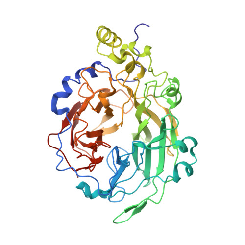
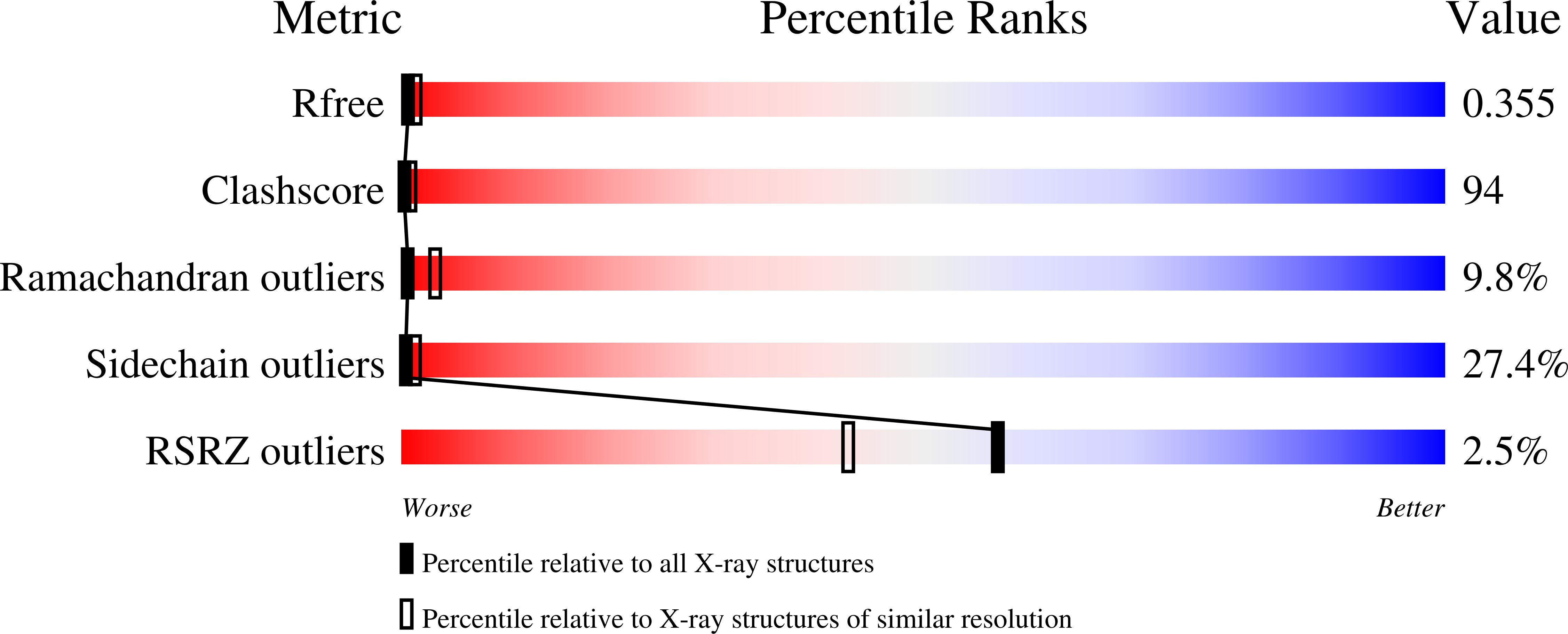
    2VDT

  • PubMed Abstract: 

    Levansucrases (LS) are fructosyltransferases (FTFs) belonging to family 68 of glycoside hydrolases (GH68) using sucrose as substrate to synthesize levan, a fructose polymer. From a multiple sequence analysis of GH68 family proteins, nine residues were selected and their role in acceptor and product specificity, as well as in biochemical Bacillus subtilis LS properties, was investigated. A product specificity modification was obtained with mutants Y429N and R433A that no longer produce levan but exclusively oligosaccharides. An effect of the mutation S164A was observed on enzyme stability and kinetic behavior; this mutation also induces a levan activation effect that enhances the reaction rate. We report the crystallographic structure of this mutant and found that S164 is an important residue to maintain the nucleophile position in the active site. We also found evidence of the important role of Y429 in acceptor specificity: this is a key residue coordinating the sucrose position in the catalytic domain-binding pocket. Some of these mutations resulted in LS with a broad range of specificities and new biochemical properties.


  • Organizational Affiliation

    Departamento de Ingeniería Celular y Biocatálisis, Instituto de Biotecnología, UNAM, Cuernavaca, Morelos 62210, Mexico.


Macromolecules
Find similar proteins by:  (by identity cutoff)  |  3D Structure
Entity ID: 1
MoleculeChains Sequence LengthOrganismDetailsImage
LEVANSUCRASE439Bacillus subtilisMutation(s): 1 
EC: 2.4.1.10
UniProt
Find proteins for P05655 (Bacillus subtilis (strain 168))
Explore P05655 
Go to UniProtKB:  P05655
Entity Groups  
Sequence Clusters30% Identity50% Identity70% Identity90% Identity95% Identity100% Identity
UniProt GroupP05655
Sequence Annotations
Expand
  • Reference Sequence
Small Molecules
Ligands 1 Unique
IDChains Name / Formula / InChI Key2D Diagram3D Interactions
CA
Query on CA

Download Ideal Coordinates CCD File 
B [auth A]CALCIUM ION
Ca
BHPQYMZQTOCNFJ-UHFFFAOYSA-N
Experimental Data & Validation

Experimental Data

  • Method: X-RAY DIFFRACTION
  • Resolution: 3.20 Å
  • R-Value Free: 0.297 
  • R-Value Work: 0.265 
  • R-Value Observed: 0.265 
  • Space Group: P 21 21 21
Unit Cell:
Length ( Å )Angle ( ˚ )
a = 50.88α = 90
b = 55.8β = 90
c = 124.6γ = 90
Software Package:
Software NamePurpose
CNSrefinement
MOSFLMdata reduction
SCALAdata scaling
PHASERphasing

Structure Validation

View Full Validation Report


Entry History 

Deposition Data

Revision History  (Full details and data files)

  • Version 1.0: 2008-07-15
    Type: Initial release
  • Version 1.1: 2011-05-08
    Changes: Version format compliance
  • Version 1.2: 2011-07-13
    Changes: Version format compliance
  • Version 1.3: 2018-06-20
    Changes: Data collection, Database references
  • Version 1.4: 2019-01-30
    Changes: Data collection, Experimental preparation
  • Version 1.5: 2023-12-13
    Changes: Data collection, Database references, Derived calculations, Other, Refinement description