A Divergent Sm Fold in Edc3 Proteins Mediates Dcp1 Binding and P-Body Targeting.
Tritschler, F., Eulalio, A., Truffault, V., Hartmann, M.D., Helms, S., Schmidt, S., Coles, M., Izaurralde, E., Weichenrieder, O.(2007) Mol Cell Biol 27: 8600
- PubMed: 17923697
- DOI: https://doi.org/10.1128/MCB.01506-07
- Primary Citation of Related Structures:
2RM4, 2VC8 - PubMed Abstract:
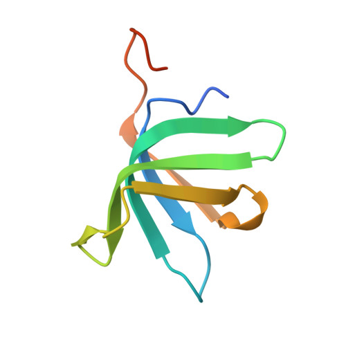
Members of the (L)Sm (Sm and Sm-like) protein family are found across all kingdoms of life and play crucial roles in RNA metabolism. The P-body component EDC3 (enhancer of decapping 3) is a divergent member of this family that functions in mRNA decapping. EDC3 is composed of a N-terminal LSm domain, a central FDF domain, and a C-terminal YjeF-N domain. We show that this modular architecture enables EDC3 to interact with multiple components of the decapping machinery, including DCP1, DCP2, and Me31B. The LSm domain mediates DCP1 binding and P-body localization. We determined the three-dimensional structures of the LSm domains of Drosophila melanogaster and human EDC3 and show that the domain adopts a divergent Sm fold that lacks the characteristic N-terminal alpha-helix and has a disrupted beta4-strand. This domain remains monomeric in solution and lacks several features that canonical (L)Sm domains require for binding RNA. The structures also revealed a conserved patch of surface residues that are required for the interaction with DCP1 but not for P-body localization. The conservation of surface and of critical structural residues indicates that LSm domains in EDC3 proteins adopt a similar fold that has separable novel functions that are absent in canonical (L)Sm proteins.
Organizational Affiliation:
Max Planck Institute for Developmental Biology, Spemannstrasse 35, D-72076 Tübingen, Germany.