2VAJ

Crystal structure of NCAM2 Ig1 (I4122 cell unit)


Experimental Data Snapshot

  • Method: X-RAY DIFFRACTION
  • Resolution: 2.70 Å
  • R-Value Free: 0.268 
  • R-Value Work: 0.227 
  • R-Value Observed: 0.229 

wwPDB Validation   3D Report Full Report


This is version 1.4 of the entry. See complete history


Literature

Crystal Structure of the Ig1 Domain of the Neural Cell Adhesion Molecule Ncam2 Displays Domain Swapping.

Rasmussen, K.K.Kulahin, N.Kristensen, O.Poulsen, J.C.Sigurskjold, B.W.Kastrup, J.S.Berezin, V.Bock, E.Walmod, P.S.Gajhede, M.

(2008) J Mol Biol 382: 1113

  • DOI: https://doi.org/10.1016/j.jmb.2008.07.084
  • Primary Citation of Related Structures:  
    2VAJ

  • PubMed Abstract: 

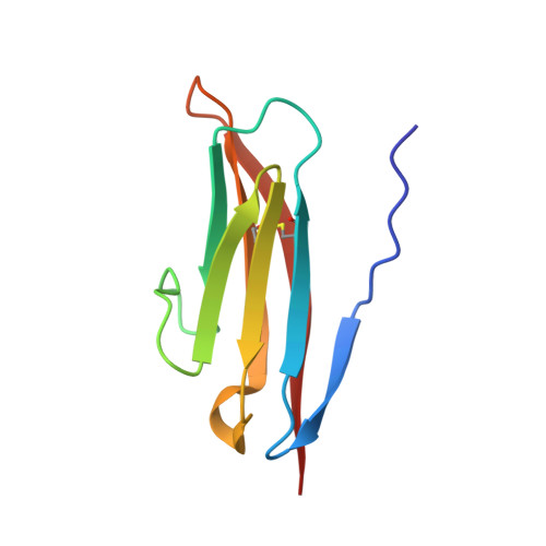
    The crystal structure of the first immunoglobulin (Ig1) domain of neural cell adhesion molecule 2 (NCAM2/OCAM/RNCAM) is presented at a resolution of 2.7 A. NCAM2 is a member of the immunoglobulin superfamily of cell adhesion molecules (IgCAMs). In the structure, two Ig domains interact by domain swapping, as the two N-terminal beta-strands are interchanged. beta-Strand swapping at the terminal domain is the accepted mechanism of homophilic interactions amongst the cadherins, another class of CAMs, but it has not been observed within the IgCAM superfamily. Gel-filtration chromatography demonstrated the ability of NCAM2 Ig1 to form dimers in solution. Taken together, these observations suggest that beta-strand swapping could have a role in the molecular mechanism of homophilic binding for NCAM2.


  • Organizational Affiliation

    Protein Laboratory, Department of Neuroscience and Pharmacology, Faculty of Health Sciences, University of Copenhagen, Blegdamsvej 3, DK-2200 Copenhagen, Denmark.


Macromolecules
Find similar proteins by:  (by identity cutoff)  |  3D Structure
Entity ID: 1
MoleculeChains Sequence LengthOrganismDetailsImage
NEURAL CELL ADHESION MOLECULE 293Homo sapiensMutation(s): 0 
UniProt & NIH Common Fund Data Resources
Find proteins for O15394 (Homo sapiens)
Explore O15394 
Go to UniProtKB:  O15394
PHAROS:  O15394
GTEx:  ENSG00000154654 
Entity Groups  
Sequence Clusters30% Identity50% Identity70% Identity90% Identity95% Identity100% Identity
UniProt GroupO15394
Sequence Annotations
Expand
  • Reference Sequence
Experimental Data & Validation

Experimental Data

  • Method: X-RAY DIFFRACTION
  • Resolution: 2.70 Å
  • R-Value Free: 0.268 
  • R-Value Work: 0.227 
  • R-Value Observed: 0.229 
  • Space Group: I 41 2 2
Unit Cell:
Length ( Å )Angle ( ˚ )
a = 47.279α = 90
b = 47.279β = 90
c = 196.727γ = 90
Software Package:
Software NamePurpose
PHENIXrefinement
MOSFLMdata reduction
SCALEPACKdata scaling
PHASERphasing

Structure Validation

View Full Validation Report


Entry History 

Deposition Data

Revision History  (Full details and data files)

  • Version 1.0: 2008-08-26
    Type: Initial release
  • Version 1.1: 2011-05-08
    Changes: Version format compliance
  • Version 1.2: 2011-07-13
    Changes: Version format compliance
  • Version 1.3: 2018-01-17
    Changes: Data collection
  • Version 1.4: 2023-12-13
    Changes: Data collection, Database references, Other, Refinement description