2V2V

IspE in complex with ligand


Experimental Data Snapshot

  • Method: X-RAY DIFFRACTION
  • Resolution: 2.40 Å
  • R-Value Free: 0.272 
  • R-Value Work: 0.217 
  • R-Value Observed: 0.220 

wwPDB Validation   3D Report Full Report


Ligand Structure Quality Assessment 


This is version 1.2 of the entry. See complete history


Literature

Synthesis and Characterization of Cytidine Derivatives that Inhibit the Kinase Ispe of the Non-Mevalonate Pathway for Isoprenoid Biosynthesis.

Crane, C.M.Hirsch, A.K.Alphey, M.S.Sgraja, T.Lauw, S.Illarionova, V.Rohdich, F.Eisenreich, W.Hunter, W.N.Bacher, A.Diederich, F.

(2008) ChemMedChem 3: 91

  • DOI: https://doi.org/10.1002/cmdc.200700208
  • Primary Citation of Related Structures:  
    2V2Q, 2V2V

  • PubMed Abstract: 

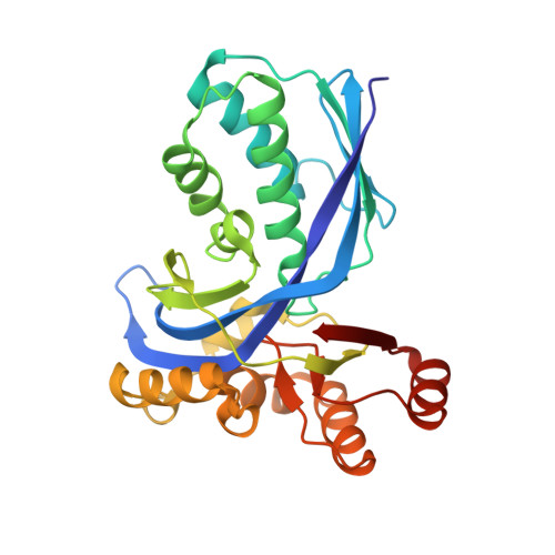
    The enzymes of the non-mevalonate pathway for isoprenoid biosynthesis are attractive targets for the development of novel drugs against malaria and tuberculosis. This pathway is used exclusively by the corresponding pathogens, but not by humans. A series of water-soluble, cytidine-based inhibitors that were originally designed for the fourth enzyme in the pathway, IspD, were shown to inhibit the subsequent enzyme, the kinase IspE (from Escherichia coli). The binding mode of the inhibitors was verified by co-crystal structure analysis, using Aquifex aeolicus IspE. The crystal structures represent the first reported example of a co-crystal structure of IspE with a synthetic ligand and confirmed that ligand binding affinity originates mainly from the interactions of the nucleobase moiety in the cytidine binding pocket of the enzyme. In contrast, the appended benzimidazole moieties of the ligands adopt various orientations in the active site and establish only poor intermolecular contacts with the protein. Defined binding sites for sulfate ions and glycerol molecules, components in the crystallization buffer, near the well-conserved ATP-binding Gly-rich loop of IspE were observed. The crystal structures of A. aeolicus IspE nicely complement the one from E. coli IspE for use in structure-based design, namely by providing invaluable structural information for the design of inhibitors targeting IspE from Mycobacterium tuberculosis and Plasmodium falciparum. Similar to the enzymes from these pathogens, A. aeolicus IspE directs the OH group of a tyrosine residue into a pocket in the active site. In the E. coli enzyme, on the other hand, this pocket is lined by phenylalanine and has a more pronounced hydrophobic character.


  • Organizational Affiliation

    Laboratorium für Organische Chemie, ETH Zürich, HCI, CH-8093 Zürich, Switzerland.


Macromolecules
Find similar proteins by:  (by identity cutoff)  |  3D Structure
Entity ID: 1
MoleculeChains Sequence LengthOrganismDetailsImage
4-DIPHOSPHOCYTIDYL-2C-METHYL-D-ERYTHRITOL KINASE
A, B
271Aquifex aeolicusMutation(s): 0 
EC: 2.7.1.148
UniProt
Find proteins for O67060 (Aquifex aeolicus (strain VF5))
Explore O67060 
Go to UniProtKB:  O67060
Entity Groups  
Sequence Clusters30% Identity50% Identity70% Identity90% Identity95% Identity100% Identity
UniProt GroupO67060
Sequence Annotations
Expand
  • Reference Sequence
Small Molecules
Ligands 4 Unique
IDChains Name / Formula / InChI Key2D Diagram3D Interactions
V12
Query on V12

Download Ideal Coordinates CCD File 
K [auth A],
T [auth B]
5'-[(1H-BENZIMIDAZOL-2-YLACETYL)AMINO]-5'-DEOXYCYTIDINE
C18 H20 N6 O5
MMCWRTZSCVUETH-GAEVZRCVSA-N
SO4
Query on SO4

Download Ideal Coordinates CCD File 
C [auth A],
D [auth A],
L [auth B],
M [auth B],
N [auth B]
SULFATE ION
O4 S
QAOWNCQODCNURD-UHFFFAOYSA-L
GOL
Query on GOL

Download Ideal Coordinates CCD File 
E [auth A],
O [auth B],
P [auth B]
GLYCEROL
C3 H8 O3
PEDCQBHIVMGVHV-UHFFFAOYSA-N
BR
Query on BR

Download Ideal Coordinates CCD File 
F [auth A]
G [auth A]
H [auth A]
I [auth A]
J [auth A]
F [auth A],
G [auth A],
H [auth A],
I [auth A],
J [auth A],
Q [auth B],
R [auth B],
S [auth B]
BROMIDE ION
Br
CPELXLSAUQHCOX-UHFFFAOYSA-M
Binding Affinity Annotations 
IDSourceBinding Affinity
V12 PDBBind:  2V2V Ki: 8.58e+4 (nM) from 1 assay(s)
Binding MOAD:  2V2V Ki: 8.58e+4 (nM) from 1 assay(s)
Experimental Data & Validation

Experimental Data

  • Method: X-RAY DIFFRACTION
  • Resolution: 2.40 Å
  • R-Value Free: 0.272 
  • R-Value Work: 0.217 
  • R-Value Observed: 0.220 
  • Space Group: P 21 3
Unit Cell:
Length ( Å )Angle ( ˚ )
a = 137.256α = 90
b = 137.256β = 90
c = 137.256γ = 90
Software Package:
Software NamePurpose
REFMACrefinement
MOSFLMdata reduction
SCALAdata scaling
REFMACphasing

Structure Validation

View Full Validation Report


Ligand Structure Quality Assessment 


Entry History 

Deposition Data

Revision History  (Full details and data files)

  • Version 1.0: 2007-06-19
    Type: Initial release
  • Version 1.1: 2011-05-08
    Changes: Version format compliance
  • Version 1.2: 2011-07-13
    Changes: Version format compliance