Evolutionary Adaptation of Human and Mouse Eye Lens Gammac-Crystallins for Solubility and Stability: Fine-Tuning of Molecular Dipoles.
Purkiss, A.G., Bateman, O.A., Wyatt, K., David, L.L., Wistow, G.J., Slingsby, C.(2007) J Mol Biol 372: 205
- PubMed: 17659303
- DOI: https://doi.org/10.1016/j.jmb.2007.06.049
- Primary Citation of Related Structures:
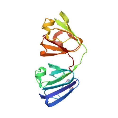
2V2U - PubMed Abstract:
The eye lens is packed with soluble crystallin proteins, providing a lifetime of transparency and light refraction. gamma-Crystallins are major components of the dense, high refractive index central regions of the lens and generally have high solubility, high stability and high levels of cysteine residues. Human gammaC belongs to a group of gamma-crystallins with a pair of cysteine residues at positions 78 and 79. Unlike other gamma-crystallins it has relatively low solubility, whereas mouse gammaC, which has the exposed C79 replaced with arginine, and a novel mouse splice variant, gammaCins, are both highly soluble. Furthermore, human gammaC is extremely stable, while the mouse orthologs are less stable. Evolutionary pressure may have favoured stability over solubility for human gammaC and the reverse for the orthologs in the mouse. Mutation of C79 to R79, in human gammaC, greatly increased solubility, however, neither form produced crystals. Remarkably, when the human gammaD R36S crystallization cataract mutation was mimicked in human gammaC-crystallin, the solubility of gammaC was dramatically increased, although it still did not crystallize. The highly soluble mouse gammaC-crystallin did crystallize. Its X-ray structure was solved and used in homology modelling of human gammaC, and its mutants C79R and R36S. The human gammaD R36S mutant was also modelled from human gammaD coordinates. Molecular dynamics simulation of the six molecules in the solution state showed that the human gammaCs differed from gammaDs in domain pairing, behaviour that correlates with interface sequence changes. When the fluctuations of the calculated molecular dipoles, for the six structures, over time were analysed, characteristic patterns for soluble gammaC and gammaD proteins were observed. Individual sequence changes that increase or decrease solubility correlated well with changes in the magnitude and direction of these dipoles. It is suggested that changes in surface residues have allowed adaptation for the differing needs of human and mouse lenses.
Organizational Affiliation:
Birkbeck College, Department of Crystallography, Institute of Structural Molecular Biology, University of London, Malet Street, London, UK.