2UZD

Crystal structure of human CDK2 complexed with a thiazolidinone inhibitor


Experimental Data Snapshot

  • Method: X-RAY DIFFRACTION
  • Resolution: 2.72 Å
  • R-Value Free: 0.269 
  • R-Value Work: 0.208 
  • R-Value Observed: 0.211 

wwPDB Validation   3D Report Full Report


Ligand Structure Quality Assessment 


This is version 1.4 of the entry. See complete history


Literature

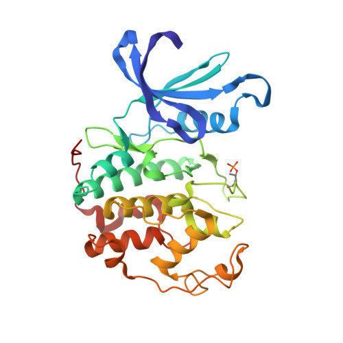
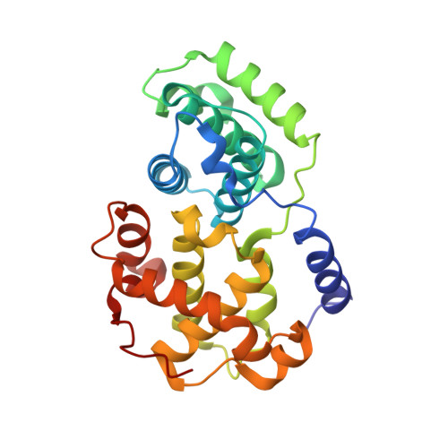
Discovery of a Potent Cdk2 Inhibitor with a Novel Binding Mode, Using Virtual Screening and Initial, Structure-Guided Lead Scoping.

Richardson, C.M.Nunns, C.L.Williamson, D.S.Parratt, M.J.Dokurno, P.Howes, R.Borgognoni, J.Drysdale, M.J.Finch, H.Hubbard, R.E.Jackson, P.S.Kierstan, P.Lentzen, G.Moore, J.D.Murray, J.B.Simmonite, H.Surgenor, A.E.Torrance, C.J.

(2007) Bioorg Med Chem Lett 17: 3880

  • DOI: https://doi.org/10.1016/j.bmcl.2007.04.110
  • Primary Citation of Related Structures:  
    2UZB, 2UZD, 2UZE, 2UZL, 2UZN, 2UZO, 2V0D

  • PubMed Abstract: 

    Virtual screening against a pCDK2/cyclin A crystal structure led to the identification of a potent and novel CDK2 inhibitor, which exhibited an unusual mode of interaction with the kinase binding motif. With the aid of X-ray crystallography and modelling, a medicinal chemistry strategy was implemented to probe the interactions seen in the crystal structure and to establish SAR. A fragment-based approach was also considered but a different, more conventional, binding mode was observed. Compound selectivity against GSK-3beta was improved using a rational design strategy, with crystallographic verification of the CDK2 binding mode.


  • Organizational Affiliation

    Vernalis (R&D) Ltd, Granta Park, Cambridge CB21 6GB, UK. c.richardson@vernalis.com


Macromolecules
Find similar proteins by:  (by identity cutoff)  |  3D Structure
Entity ID: 1
MoleculeChains Sequence LengthOrganismDetailsImage
CELL DIVISION PROTEIN KINASE 2
A, C
298Homo sapiensMutation(s): 0 
EC: 2.7.11.22 (PDB Primary Data), 2.7.1.37 (PDB Primary Data)
UniProt & NIH Common Fund Data Resources
Find proteins for P24941 (Homo sapiens)
Explore P24941 
Go to UniProtKB:  P24941
PHAROS:  P24941
GTEx:  ENSG00000123374 
Entity Groups  
Sequence Clusters30% Identity50% Identity70% Identity90% Identity95% Identity100% Identity
UniProt GroupP24941
Sequence Annotations
Expand
  • Reference Sequence
Find similar proteins by:  (by identity cutoff)  |  3D Structure
Entity ID: 2
MoleculeChains Sequence LengthOrganismDetailsImage
CYCLIN A2
B, D
258Homo sapiensMutation(s): 0 
UniProt & NIH Common Fund Data Resources
Find proteins for P20248 (Homo sapiens)
Explore P20248 
Go to UniProtKB:  P20248
PHAROS:  P20248
GTEx:  ENSG00000145386 
Entity Groups  
Sequence Clusters30% Identity50% Identity70% Identity90% Identity95% Identity100% Identity
UniProt GroupP20248
Sequence Annotations
Expand
  • Reference Sequence
Small Molecules
Ligands 1 Unique
IDChains Name / Formula / InChI Key2D Diagram3D Interactions
C85
Query on C85

Download Ideal Coordinates CCD File 
E [auth A],
F [auth C]
4-{5-[(Z)-(2-IMINO-4-OXO-1,3-THIAZOLIDIN-5-YLIDENE)METHYL]FURAN-2-YL}BENZENESULFONAMIDE
C14 H11 N3 O4 S2
BKUMVXIXUVYKDQ-GHXNOFRVSA-N
Modified Residues  1 Unique
IDChains TypeFormula2D DiagramParent
TPO
Query on TPO
A, C
L-PEPTIDE LINKINGC4 H10 N O6 PTHR
Binding Affinity Annotations 
IDSourceBinding Affinity
C85 PDBBind:  2UZD IC50: 180 (nM) from 1 assay(s)
BindingDB:  2UZD IC50: 180 (nM) from 1 assay(s)
Experimental Data & Validation

Experimental Data

  • Method: X-RAY DIFFRACTION
  • Resolution: 2.72 Å
  • R-Value Free: 0.269 
  • R-Value Work: 0.208 
  • R-Value Observed: 0.211 
  • Space Group: P 21 21 2
Unit Cell:
Length ( Å )Angle ( ˚ )
a = 160.559α = 90
b = 160.292β = 90
c = 66.509γ = 90
Software Package:
Software NamePurpose
REFMACrefinement
DENZOdata reduction
SCALEPACKdata scaling

Structure Validation

View Full Validation Report


Ligand Structure Quality Assessment 


Entry History 

Deposition Data

Revision History  (Full details and data files)

  • Version 1.0: 2007-06-26
    Type: Initial release
  • Version 1.1: 2011-05-08
    Changes: Version format compliance
  • Version 1.2: 2011-07-13
    Changes: Version format compliance
  • Version 1.3: 2019-03-06
    Changes: Data collection, Derived calculations, Experimental preparation, Other
  • Version 1.4: 2019-04-03
    Changes: Data collection, Experimental preparation