2UV1

Hexagonal crystal form of GamS from bacteriophage lambda.


Experimental Data Snapshot

  • Method: X-RAY DIFFRACTION
  • Resolution: 2.60 Å
  • R-Value Free: 0.300 
  • R-Value Work: 0.235 
  • R-Value Observed: 0.239 

wwPDB Validation   3D Report Full Report


This is version 1.3 of the entry. See complete history


Literature

The Crystal Structure of Lambda-Gam Protein Suggests a Model for Recbcd Inhibition.

Court, R.I.Cook, N.Saikrishnan, K.Wigley, D.B.

(2007) J Mol Biol 371: 25

  • DOI: https://doi.org/10.1016/j.jmb.2007.05.037
  • Primary Citation of Related Structures:  
    2UUZ, 2UV1

  • PubMed Abstract: 

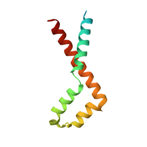
    In Escherichia coli, RecBCD processes double-stranded DNA breaks during the initial stages of homologous recombination. RecBCD contains helicase and nuclease activities, and unwinds and digests the blunt-ended DNA until a specific eight-nucleotide sequence, Chi, is encountered. Chi modulates the nuclease activity of RecBCD and results in a resected DNA end, which is a substrate for RecA during subsequent steps in recombination. RecBCD also acts as a defence mechanism against bacteriophage infection by digesting linear viral DNA present during virus replication or resulting from the action of restriction endonucleases. To avoid this fate, bacteriophage lambda encodes the gene Gam whose product is an inhibitor of RecBCD. Gam has been shown to bind to RecBCD and inhibit its helicase and nuclease activities. We show that Gam inhibits RecBCD by preventing it from binding DNA. We have solved the crystal structure of Gam from two different crystal forms. Using the published crystal structure of RecBCD in complex with DNA we suggest models for the molecular mechanism of Gam-mediated inhibition of RecBCD. We also propose that Gam could be a mimetic of single-stranded, and perhaps also double-stranded, DNA.


  • Organizational Affiliation

    Cancer Research UK Clare Hall Laboratories, Blanche Lane, South Mimms, Hertfordshire EN6 3LD, UK.


Macromolecules
Find similar proteins by:  (by identity cutoff)  |  3D Structure
Entity ID: 1
MoleculeChains Sequence LengthOrganismDetailsImage
HOST-NUCLEASE INHIBITOR PROTEIN GAM
A, B
99Lambdavirus lambdaMutation(s): 0 
UniProt
Find proteins for P03702 (Escherichia phage lambda)
Explore P03702 
Go to UniProtKB:  P03702
Entity Groups  
Sequence Clusters30% Identity50% Identity70% Identity90% Identity95% Identity100% Identity
UniProt GroupP03702
Sequence Annotations
Expand
  • Reference Sequence
Experimental Data & Validation

Experimental Data

  • Method: X-RAY DIFFRACTION
  • Resolution: 2.60 Å
  • R-Value Free: 0.300 
  • R-Value Work: 0.235 
  • R-Value Observed: 0.239 
  • Space Group: P 6 2 2
Unit Cell:
Length ( Å )Angle ( ˚ )
a = 84.479α = 90
b = 84.479β = 90
c = 105.26γ = 120
Software Package:
Software NamePurpose
REFMACrefinement
MOSFLMdata reduction
SCALAdata scaling
PHASERphasing

Structure Validation

View Full Validation Report


Entry History 

Deposition Data

Revision History  (Full details and data files)

  • Version 1.0: 2007-06-12
    Type: Initial release
  • Version 1.1: 2011-05-08
    Changes: Version format compliance
  • Version 1.2: 2011-07-13
    Changes: Version format compliance
  • Version 1.3: 2023-12-13
    Changes: Data collection, Database references, Other, Refinement description