2TRM

THE THREE-DIMENSIONAL STRUCTURE OF ASN102 MUTANT OF TRYPSIN. ROLE OF ASP102 IN SERINE PROTEASE CATALYSIS


Experimental Data Snapshot

  • Method: X-RAY DIFFRACTION
  • Resolution: 2.80 Å
  • R-Value Observed: 0.157 

wwPDB Validation   3D Report Full Report


This is version 1.3 of the entry. See complete history


Literature

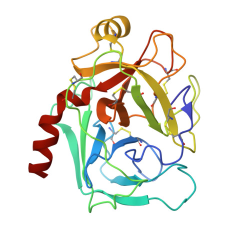
The three-dimensional structure of Asn102 mutant of trypsin: role of Asp102 in serine protease catalysis.

Sprang, S.Standing, T.Fletterick, R.J.Stroud, R.M.Finer-Moore, J.Xuong, N.H.Hamlin, R.Rutter, W.J.Craik, C.S.

(1987) Science 237: 905-909

  • DOI: https://doi.org/10.1126/science.3112942
  • Primary Citation of Related Structures:  
    1TRM, 2TRM

  • PubMed Abstract: 

    The structure of the Asn102 mutant of trypsin was determined in order to distinguish whether the reduced activity of the mutant at neutral pH results from an altered active site conformation or from an inability to stabilize a positive charge on the active site histidine. The active site structure of the Asn102 mutant of trypsin is identical to the native enzyme with respect to the specificity pocket, the oxyanion hole, and the orientation of the nucleophilic serine. The observed decrease in rate results from the loss of nucleophilicity of the active site serine. This decreased nucleophilicity may result from stabilization of a His57 tautomer that is unable to accept the serine hydroxyl proton.


Macromolecules
Find similar proteins by:  (by identity cutoff)  |  3D Structure
Entity ID: 1
MoleculeChains Sequence LengthOrganismDetailsImage
TRYPSIN223Rattus norvegicusMutation(s): 0 
EC: 3.4.21.4
UniProt
Find proteins for P00763 (Rattus norvegicus)
Explore P00763 
Go to UniProtKB:  P00763
Entity Groups  
Sequence Clusters30% Identity50% Identity70% Identity90% Identity95% Identity100% Identity
UniProt GroupP00763
Sequence Annotations
Expand
  • Reference Sequence
Small Molecules
Ligands 2 Unique
IDChains Name / Formula / InChI Key2D Diagram3D Interactions
BEN
Query on BEN

Download Ideal Coordinates CCD File 
C [auth A]BENZAMIDINE
C7 H8 N2
PXXJHWLDUBFPOL-UHFFFAOYSA-N
CA
Query on CA

Download Ideal Coordinates CCD File 
B [auth A]CALCIUM ION
Ca
BHPQYMZQTOCNFJ-UHFFFAOYSA-N
Experimental Data & Validation

Experimental Data

  • Method: X-RAY DIFFRACTION
  • Resolution: 2.80 Å
  • R-Value Observed: 0.157 
  • Space Group: I 2 3
Unit Cell:
Length ( Å )Angle ( ˚ )
a = 124.38α = 90
b = 124.38β = 90
c = 124.38γ = 90
Software Package:
Software NamePurpose
PROLSQrefinement

Structure Validation

View Full Validation Report


Entry History 

Deposition Data

Revision History  (Full details and data files)

  • Version 1.0: 1988-07-16
    Type: Initial release
  • Version 1.1: 2008-03-03
    Changes: Version format compliance
  • Version 1.2: 2011-07-13
    Changes: Version format compliance
  • Version 1.3: 2017-11-29
    Changes: Derived calculations, Other