2SPC

CRYSTAL STRUCTURE OF THE REPETITIVE SEGMENTS OF SPECTRIN


Experimental Data Snapshot

  • Method: X-RAY DIFFRACTION
  • Resolution: 1.80 Å
  • R-Value Work: 0.203 
  • R-Value Observed: 0.203 

wwPDB Validation   3D Report Full Report


This is version 1.6 of the entry. See complete history


Literature

Crystal structure of the repetitive segments of spectrin.

Yan, Y.Winograd, E.Viel, A.Cronin, T.Harrison, S.C.Branton, D.

(1993) Science 262: 2027-2030

  • DOI: https://doi.org/10.1126/science.8266097
  • Primary Citation of Related Structures:  
    2SPC

  • PubMed Abstract: 

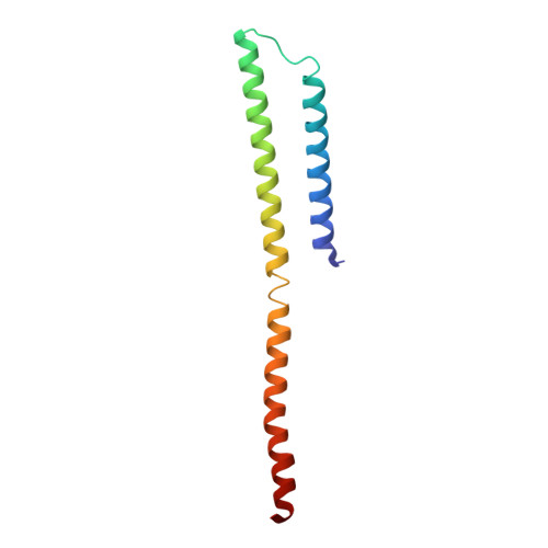
    The elongated proteins of the spectrin family (dystrophin, alpha-actinin, and spectrin) contain tandemly repeated segments and form resilient cellular meshworks by cross-linking actin filaments. The structure of one of the repetitive segments of alpha-spectrin was determined at a 1.8 angstrom resolution. A segment consists of a three-helix bundle. A model of the interface between two tandem segments suggests that hydrophobic interactions between segments may constrain intersegment flexibility. The helix side chain interactions explain how mutations that are known to produce hemolytic anemias disrupt spectrin associations that sustain the integrity of the erythrocyte membrane.


  • Organizational Affiliation

    Department of Biochemistry, Harvard University, Cambridge, MA 02138.


Macromolecules
Find similar proteins by:  (by identity cutoff)  |  3D Structure
Entity ID: 1
MoleculeChains Sequence LengthOrganismDetailsImage
SPECTRIN
A, B
107Drosophila melanogasterMutation(s): 0 
UniProt
Find proteins for P13395 (Drosophila melanogaster)
Explore P13395 
Go to UniProtKB:  P13395
Entity Groups  
Sequence Clusters30% Identity50% Identity70% Identity90% Identity95% Identity100% Identity
UniProt GroupP13395
Sequence Annotations
Expand
  • Reference Sequence
Experimental Data & Validation

Experimental Data

  • Method: X-RAY DIFFRACTION
  • Resolution: 1.80 Å
  • R-Value Work: 0.203 
  • R-Value Observed: 0.203 
  • Space Group: P 21 21 2
Unit Cell:
Length ( Å )Angle ( ˚ )
a = 46.8α = 90
b = 47.22β = 90
c = 104.43γ = 90
Software Package:
Software NamePurpose
X-PLORmodel building
TNTrefinement
X-PLORrefinement
X-PLORphasing

Structure Validation

View Full Validation Report


Entry History 

Deposition Data

Revision History  (Full details and data files)

  • Version 1.0: 1994-05-31
    Type: Initial release
  • Version 1.1: 2008-03-25
    Changes: Version format compliance
  • Version 1.2: 2011-07-13
    Changes: Version format compliance
  • Version 1.3: 2011-11-16
    Changes: Atomic model
  • Version 1.4: 2019-07-17
    Changes: Data collection, Other, Refinement description
  • Version 1.5: 2019-08-14
    Changes: Data collection, Refinement description
  • Version 1.6: 2024-02-21
    Changes: Data collection, Database references