2SKC

PYRIDOXAL PHOSPHORYLASE B IN COMPLEX WITH FLUOROPHOSPHATE, GLUCOSE AND INOSINE-5'-MONOPHOSPHATE


Experimental Data Snapshot

  • Method: X-RAY DIFFRACTION
  • Resolution: 2.40 Å
  • R-Value Free: 0.217 
  • R-Value Work: 0.167 
  • R-Value Observed: 0.167 

wwPDB Validation   3D Report Full Report


This is version 1.4 of the entry. See complete history


Literature

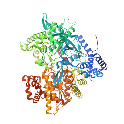
Activator anion binding site in pyridoxal phosphorylase b: the binding of phosphite, phosphate, and fluorophosphate in the crystal.

Oikonomakos, N.G.Zographos, S.E.Tsitsanou, K.E.Johnson, L.N.Acharya, K.R.

(1996) Protein Sci 5: 2416-2428

  • DOI: https://doi.org/10.1002/pro.5560051204
  • Primary Citation of Related Structures:  
    2SKC, 2SKD, 2SKE

  • PubMed Abstract: 

    It has been established that phosphate analogues can activate glycogen phosphorylase reconstituted with pyridoxal in place of the natural cofactor pyridoxal 5'-phosphate (Change YC. McCalmont T, Graves DJ. 1983. Biochemistry 22:4987-4993). Pyridoxal phosphorylase b has been studied by kinetic, ultracentrifugation, and X-ray crystallographic experiments. In solution, the catalytically active species of pyridoxal phosphorylase b adopts a conformation that is more R-state-like than that of native phosphorylase b, but an inactive dimeric species of the enzyme can be stabilized by activator phosphite in combination with the T-state inhibitor glucose. Co-crystals of pyridoxal phosphorylase b complexed with either phosphite, phosphate, or fluorophosphate, the inhibitor glucose, and the weak activator IMP were grown in space group P4(3)2(1)2, with native-like unit cell dimensions, and the structures of the complexes have been refined to give crystallographic R factors of 18.5-19.2%, for data between 8 and 2.4 A resolution. The anions bind tightly at the catalytic site in a similar but not identical position to that occupied by the cofactor 5'-phosphate group in the native enzyme (phosphorus to phosphorus atoms distance = 1.2 A). The structural results show that the structures of the pyridoxal phosphorylase b-anion-glucose-IMP complexes are overall similar to the glucose complex of native T-state phosphorylase b. Structural comparisons suggest that the bound anions, in the position observed in the crystal, might have a structural role for effective catalysis.


  • Organizational Affiliation

    Institute of Biological Research and Biotechnology, National Hellenic Research Foundation, Athens, Greece. nikos@apollon.servicenet.ariadne-t.gr


Macromolecules
Find similar proteins by:  (by identity cutoff)  |  3D Structure
Entity ID: 1
MoleculeChains Sequence LengthOrganismDetailsImage
PYRIDOXAL PHOSPHORYLASE B842Oryctolagus cuniculusMutation(s): 0 
EC: 2.4.1.1
UniProt
Find proteins for P00489 (Oryctolagus cuniculus)
Explore P00489 
Go to UniProtKB:  P00489
Entity Groups  
Sequence Clusters30% Identity50% Identity70% Identity90% Identity95% Identity100% Identity
UniProt GroupP00489
Sequence Annotations
Expand
  • Reference Sequence
Small Molecules
Ligands 4 Unique
IDChains Name / Formula / InChI Key2D Diagram3D Interactions
IMP
Query on IMP

Download Ideal Coordinates CCD File 
E [auth A]INOSINIC ACID
C10 H13 N4 O8 P
GRSZFWQUAKGDAV-KQYNXXCUSA-N
PLP
Query on PLP

Download Ideal Coordinates CCD File 
C [auth A]PYRIDOXAL-5'-PHOSPHATE
C8 H10 N O6 P
NGVDGCNFYWLIFO-UHFFFAOYSA-N
GLC
Query on GLC

Download Ideal Coordinates CCD File 
B [auth A]alpha-D-glucopyranose
C6 H12 O6
WQZGKKKJIJFFOK-DVKNGEFBSA-N
FPO
Query on FPO

Download Ideal Coordinates CCD File 
D [auth A]FLUORO-PHOSPHITE ION
F O3 P
DWYMPOCYEZONEA-UHFFFAOYSA-L
Experimental Data & Validation

Experimental Data

  • Method: X-RAY DIFFRACTION
  • Resolution: 2.40 Å
  • R-Value Free: 0.217 
  • R-Value Work: 0.167 
  • R-Value Observed: 0.167 
  • Space Group: P 43 21 2
Unit Cell:
Length ( Å )Angle ( ˚ )
a = 128.5α = 90
b = 128.5β = 90
c = 116.3γ = 90
Software Package:
Software NamePurpose
X-PLORmodel building
X-PLORrefinement
MOSCOdata reduction
CCP4data scaling
X-PLORphasing

Structure Validation

View Full Validation Report


Entry History 

Deposition Data

Revision History  (Full details and data files)

  • Version 1.0: 1998-12-16
    Type: Initial release
  • Version 1.1: 2008-04-26
    Changes: Version format compliance
  • Version 1.2: 2011-07-13
    Changes: Derived calculations, Version format compliance
  • Version 1.3: 2020-07-29
    Type: Remediation
    Reason: Carbohydrate remediation
    Changes: Data collection, Database references, Derived calculations, Structure summary
  • Version 1.4: 2023-08-30
    Changes: Data collection, Database references, Refinement description, Structure summary