2RGS

FC-fragment of monoclonal antibody IGG2B from Mus musculus


Experimental Data Snapshot

  • Method: X-RAY DIFFRACTION
  • Resolution: 2.13 Å
  • R-Value Free: 0.213 
  • R-Value Work: 0.201 
  • R-Value Observed: 0.201 

wwPDB Validation   3D Report Full Report


This is version 2.1 of the entry. See complete history


Literature

New insights into intra- and intermolecular interactions of immunoglobulins: crystal structure of mouse IgG2b-Fc at 2.1-A resolution

Kolenko, P.Dohnalek, J.Duskova, J.Skalova, T.Collard, R.Hasek, J.

(2008) Immunology 126: 378-385

  • DOI: https://doi.org/10.1111/j.1365-2567.2008.02904.x
  • Primary Citation of Related Structures:  
    2RGS

  • PubMed Abstract: 

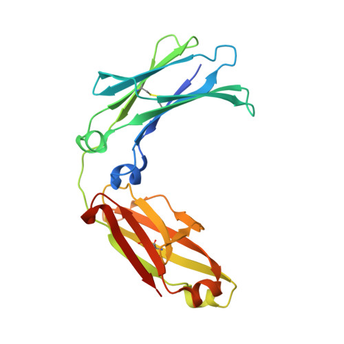
    The structure of the Fc fragment of monoclonal antibody IgG2b from hybridom M75 of Mus musculus has been determined by single crystal X-ray diffraction. This is the first report of the structure of the murine immunoglobulin isotype IgG2b. The structure refined at 2.1 A resolution provides more detailed structural information about native oligosaccharides than was previously available. High-quality Fourier maps provide a clear identification of alpha-l-fucose with partial occupancy in the first branch of the antennary oligosaccharides. A unique Fc:Fc interaction was observed at the C(H)2-C(H)3 interface.


  • Organizational Affiliation

    Department of Structure Analysis, Institute of Macromolecular Chemistry AS CR, Praha, Czech Republic. hasek@imc.cas.cz


Macromolecules
Find similar proteins by:  (by identity cutoff)  |  3D Structure
Entity ID: 1
MoleculeChains Sequence LengthOrganismDetailsImage
Ig gamma-2B heavy chain
A, B
218Mus musculusMutation(s): 0 
UniProt
Find proteins for P01867 (Mus musculus)
Explore P01867 
Go to UniProtKB:  P01867
Entity Groups  
Sequence Clusters30% Identity50% Identity70% Identity90% Identity95% Identity100% Identity
UniProt GroupP01867
Sequence Annotations
Expand
  • Reference Sequence
Oligosaccharides

Help

Entity ID: 2
MoleculeChains Length2D Diagram Glycosylation3D Interactions
beta-D-galactopyranose-(1-4)-2-acetamido-2-deoxy-beta-D-glucopyranose-(1-2)-alpha-D-mannopyranose-(1-6)-[2-acetamido-2-deoxy-beta-D-glucopyranose-(1-2)-alpha-D-mannopyranose-(1-3)]alpha-D-mannopyranose-(1-4)-2-acetamido-2-deoxy-beta-D-glucopyranose-(1-4)-[alpha-L-fucopyranose-(1-6)]2-acetamido-2-deoxy-beta-D-glucopyranose
C, D
9N-Glycosylation
Glycosylation Resources
GlyTouCan:  G58978IW
GlyCosmos:  G58978IW
GlyGen:  G58978IW
Experimental Data & Validation

Experimental Data

  • Method: X-RAY DIFFRACTION
  • Resolution: 2.13 Å
  • R-Value Free: 0.213 
  • R-Value Work: 0.201 
  • R-Value Observed: 0.201 
  • Space Group: C 1 2 1
Unit Cell:
Length ( Å )Angle ( ˚ )
a = 138.946α = 90
b = 63.956β = 103.83
c = 72.624γ = 90
Software Package:
Software NamePurpose
ADSCdata collection
AMoREphasing
REFMACrefinement
DENZOdata reduction
SCALEPACKdata scaling

Structure Validation

View Full Validation Report


Entry History 

Deposition Data

Revision History  (Full details and data files)

  • Version 1.0: 2008-10-07
    Type: Initial release
  • Version 1.1: 2011-07-13
    Changes: Non-polymer description, Version format compliance
  • Version 2.0: 2020-07-29
    Type: Remediation
    Reason: Carbohydrate remediation
    Changes: Advisory, Atomic model, Data collection, Derived calculations, Structure summary
  • Version 2.1: 2023-08-30
    Changes: Data collection, Database references, Refinement description, Structure summary