2QS4

Crystal structure of the GluR5 ligand binding core dimer in complex with LY466195 at 1.58 Angstroms resolution


Experimental Data Snapshot

  • Method: X-RAY DIFFRACTION
  • Resolution: 1.58 Å
  • R-Value Free: 0.199 
  • R-Value Work: 0.158 
  • R-Value Observed: 0.160 

wwPDB Validation   3D Report Full Report


Ligand Structure Quality Assessment 


This is version 1.6 of the entry. See complete history


Literature

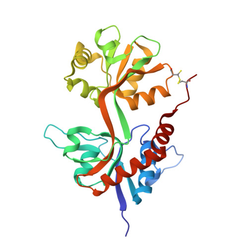
Binding site and ligand flexibility revealed by high resolution crystal structures of GluK1 competitive antagonists.

Alushin, G.M.Jane, D.Mayer, M.L.

(2011) Neuropharmacology 60: 126-134

  • DOI: https://doi.org/10.1016/j.neuropharm.2010.06.002
  • Primary Citation of Related Structures:  
    2QS1, 2QS2, 2QS4

  • PubMed Abstract: 

    The availability of crystal structures for the ligand binding domains of ionotropic glutamate receptors, combined with their key role in synaptic function in the normal and diseased brain, offers a unique selection of targets for pharmaceutical research compared to other drug targets for which the atomic structure of the ligand binding site is not known. Currently only a few antagonist structures have been solved, and these reveal ligand specific conformational changes that hinder rational drug design. Here we report high resolution crystal structures for three kainate receptor GluK1 antagonist complexes which reveal new and unexpected modes of binding, highlighting the continued need for experimentally determined receptor-ligand complexes.


  • Organizational Affiliation

    Laboratory of Cellular and Molecular Neurophysiology, Porter Neuroscience Research Center, NICHD, NIH, DHHS, Bethesda, MD 20892, USA.


Macromolecules
Find similar proteins by:  (by identity cutoff)  |  3D Structure
Entity ID: 1
MoleculeChains Sequence LengthOrganismDetailsImage
Glutamate receptor, ionotropic kainate 1
A, B, C, D
258Rattus norvegicusMutation(s): 1 
Gene Names: Grik1Glur5
UniProt
Find proteins for P22756 (Rattus norvegicus)
Explore P22756 
Go to UniProtKB:  P22756
Entity Groups  
Sequence Clusters30% Identity50% Identity70% Identity90% Identity95% Identity100% Identity
UniProt GroupP22756
Sequence Annotations
Expand
  • Reference Sequence
Small Molecules
Ligands 3 Unique
IDChains Name / Formula / InChI Key2D Diagram3D Interactions
LY5
Query on LY5

Download Ideal Coordinates CCD File 
F [auth A],
G [auth B],
I [auth C],
L [auth D]
(3S,4aR,6S,8aR)-6-{[(2S)-2-carboxy-4,4-difluoropyrrolidin-1-yl]methyl}decahydroisoquinoline-3-carboxylic acid
C16 H24 F2 N2 O4
OXQXJYQSWZFDBB-WJTVCTBASA-N
GOL
Query on GOL

Download Ideal Coordinates CCD File 
H [auth B],
J [auth C]
GLYCEROL
C3 H8 O3
PEDCQBHIVMGVHV-UHFFFAOYSA-N
NH4
Query on NH4

Download Ideal Coordinates CCD File 
E [auth A],
K [auth D]
AMMONIUM ION
H4 N
QGZKDVFQNNGYKY-UHFFFAOYSA-O
Binding Affinity Annotations 
IDSourceBinding Affinity
LY5 BindingDB:  2QS4 Ki: min: 50, max: 50 (nM) from 2 assay(s)
Binding MOAD:  2QS4 Kd: 38 (nM) from 1 assay(s)
Experimental Data & Validation

Experimental Data

  • Method: X-RAY DIFFRACTION
  • Resolution: 1.58 Å
  • R-Value Free: 0.199 
  • R-Value Work: 0.158 
  • R-Value Observed: 0.160 
  • Space Group: H 3
Unit Cell:
Length ( Å )Angle ( ˚ )
a = 89.241α = 90
b = 89.241β = 90
c = 330.365γ = 120
Software Package:
Software NamePurpose
REFMACrefinement
SERGUIdata collection
HKL-2000data reduction
HKL-2000data scaling
PHASERphasing

Structure Validation

View Full Validation Report


Ligand Structure Quality Assessment 


Entry History 

Deposition Data

Revision History  (Full details and data files)

  • Version 1.0: 2008-08-05
    Type: Initial release
  • Version 1.1: 2011-07-13
    Changes: Advisory, Version format compliance
  • Version 1.2: 2012-08-22
    Changes: Database references
  • Version 1.3: 2015-08-05
    Changes: Non-polymer description
  • Version 1.4: 2017-08-02
    Changes: Source and taxonomy
  • Version 1.5: 2021-10-20
    Changes: Database references, Derived calculations
  • Version 1.6: 2023-08-30
    Changes: Data collection, Refinement description