An alternative excited-state proton transfer pathway in green fluorescent protein variant S205V.
Shu, X., Leiderman, P., Gepshtein, R., Smith, N.R., Kallio, K., Huppert, D., Remington, S.J.(2007) Protein Sci 16: 2703-2710
- PubMed: 17965188
- DOI: https://doi.org/10.1110/ps.073112007
- Primary Citation of Related Structures:
2QLE - PubMed Abstract:
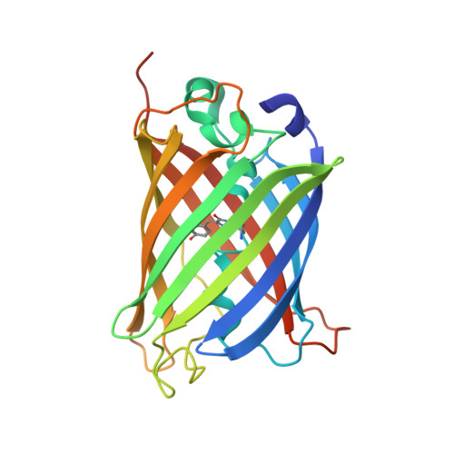
Wild-type green fluorescent protein (wt-GFP) has a prominent absorbance band centered at approximately 395 nm, attributed to the neutral chromophore form. The green emission arising upon excitation of this band results from excited-state proton transfer (ESPT) from the chromophore hydroxyl, through a hydrogen-bond network proposed to consist of a water molecule and Ser205, to Glu222. Although evidence for Glu222 as a terminal proton acceptor has already been obtained, no evidence for the participation of Ser205 in the proton transfer process exists. To examine the role of Ser205 in the proton transfer, we mutated Ser205 to valine. However, the derived GFP variant S205V, upon excitation at 400 nm, still produces green fluorescence. Time-resolved emission spectroscopy suggests that ESPT contributes to the green fluorescence, and that the proton transfer takes place approximately 30 times more slowly than in wt-GFP. The crystal structure of S205V reveals rearrangement of Glu222 and Thr203, forming a new hydrogen-bonding network. We propose this network to be an alternative ESPT pathway with distinctive features that explain the significantly slowed rate of proton transfer. In support of this proposal, the double mutant S205V/T203V is shown to be a novel blue fluorescent protein containing a tyrosine-based chromophore, yet is incapable of ESPT. The results have implications for the detailed mechanism of ESPT and the photocycle of wt-GFP, in particular for the structures of spectroscopically identified intermediates in the cycle.
Organizational Affiliation:
Institute of Molecular Biology and Department of Physics, University of Oregon, Eugene, Oregon 97403-1229, USA.