2QKU

The 5th PDZ Domain of InaD in 10mM DTT


Experimental Data Snapshot

  • Method: X-RAY DIFFRACTION
  • Resolution: 2.20 Å
  • R-Value Free: 0.231 
  • R-Value Work: 0.206 
  • R-Value Observed: 0.206 

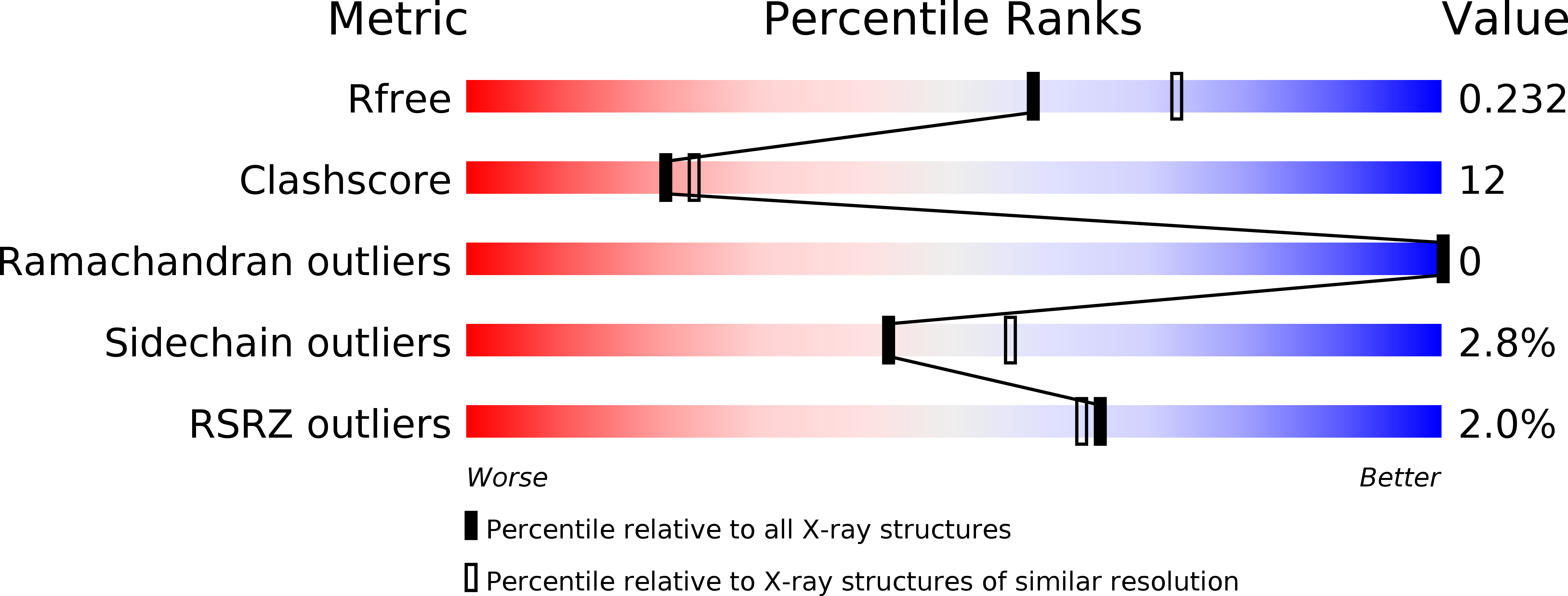
wwPDB Validation   3D Report Full Report


This is version 1.1 of the entry. See complete history


Literature

Dynamic Scaffolding in a G Protein-Coupled Signaling System.

Mishra, P.Socolich, M.Wall, M.A.Graves, J.Wang, Z.Ranganathan, R.

(2007) Cell 131: 80-92

  • DOI: https://doi.org/10.1016/j.cell.2007.07.037
  • Primary Citation of Related Structures:  
    2QKT, 2QKU, 2QKV

  • PubMed Abstract: 

    The INAD scaffold organizes a multiprotein complex that is essential for proper visual signaling in Drosophila photoreceptor cells. Here we show that one of the INAD PDZ domains (PDZ5) exists in a redox-dependent equilibrium between two conformations--a reduced form that is similar to the structure of other PDZ domains, and an oxidized form in which the ligand-binding site is distorted through formation of a strong intramolecular disulfide bond. We demonstrate transient light-dependent formation of this disulfide bond in vivo and find that transgenic flies expressing a mutant INAD in which PDZ5 is locked in the reduced state display severe defects in termination of visual responses and visually mediated reflex behavior. These studies demonstrate a conformational switch mechanism for PDZ domain function and suggest that INAD behaves more like a dynamic machine rather than a passive scaffold, regulating signal transduction at the millisecond timescale through cycles of conformational change.


  • Organizational Affiliation

    Howard Hughes Medical Institute, University of Texas Southwestern Medical Center, Dallas, TX 75390-9050, USA.


Macromolecules
Find similar proteins by:  (by identity cutoff)  |  3D Structure
Entity ID: 1
MoleculeChains Sequence LengthOrganismDetailsImage
Inactivation-no-after-potential D protein
A, B, C
90Drosophila melanogasterMutation(s): 0 
Gene Names: inaD
UniProt
Find proteins for Q24008 (Drosophila melanogaster)
Explore Q24008 
Go to UniProtKB:  Q24008
Entity Groups  
Sequence Clusters30% Identity50% Identity70% Identity90% Identity95% Identity100% Identity
UniProt GroupQ24008
Sequence Annotations
Expand
  • Reference Sequence
Small Molecules
Modified Residues  1 Unique
IDChains TypeFormula2D DiagramParent
MSE
Query on MSE
A, B, C
L-PEPTIDE LINKINGC5 H11 N O2 SeMET
Experimental Data & Validation

Experimental Data

  • Method: X-RAY DIFFRACTION
  • Resolution: 2.20 Å
  • R-Value Free: 0.231 
  • R-Value Work: 0.206 
  • R-Value Observed: 0.206 
  • Space Group: P 43 2 2
Unit Cell:
Length ( Å )Angle ( ˚ )
a = 67.7α = 90
b = 67.7β = 90
c = 162.5γ = 90
Software Package:
Software NamePurpose
CNSrefinement
PDB_EXTRACTdata extraction
HKL-2000data collection
HKL-2000data reduction
SCALEPACKdata scaling
CNSphasing

Structure Validation

View Full Validation Report


Entry History 

Deposition Data

Revision History  (Full details and data files)

  • Version 1.0: 2007-11-06
    Type: Initial release
  • Version 1.1: 2011-07-13
    Changes: Non-polymer description, Version format compliance