2QI9

ABC-transporter BtuCD in complex with its periplasmic binding protein BtuF


Experimental Data Snapshot

  • Method: X-RAY DIFFRACTION
  • Resolution: 2.60 Å
  • R-Value Free: 0.280 
  • R-Value Work: 0.262 

wwPDB Validation   3D Report Full Report


This is version 1.3 of the entry. See complete history


Literature

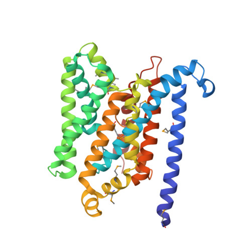
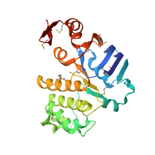
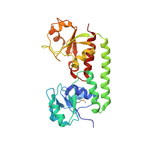
Asymmetry in the structure of the ABC transporter-binding protein complex BtuCD-BtuF.

Hvorup, R.N.Goetz, B.A.Niederer, M.Hollenstein, K.Perozo, E.Locher, K.P.

(2007) Science 317: 1387-1390

  • DOI: https://doi.org/10.1126/science.1145950
  • Primary Citation of Related Structures:  
    2QI9

  • PubMed Abstract: 

    BtuCD is an adenosine triphosphate-binding cassette (ABC) transporter that translocates vitamin B12 from the periplasmic binding protein BtuF into the cytoplasm of Escherichia coli. The 2.6 angstrom crystal structure of a complex BtuCD-F reveals substantial conformational changes as compared with the previously reported structures of BtuCD and BtuF. The lobes of BtuF are spread apart, and B12 is displaced from the binding pocket. The transmembrane BtuC subunits reveal two distinct conformations, and the translocation pathway is closed to both sides of the membrane. Electron paramagnetic resonance spectra of spin-labeled cysteine mutants reconstituted in proteoliposomes are consistent with the conformation of BtuCD-F that was observed in the crystal structure. A comparison with BtuCD and the homologous HI1470/71 protein suggests that the structure of BtuCD-F may reflect a posttranslocation intermediate.


  • Organizational Affiliation

    Institute of Molecular Biology and Biophysics, ETH Zurich, HPK D14.3, 8093 Zurich, Switzerland.


Macromolecules
Find similar proteins by:  (by identity cutoff)  |  3D Structure
Entity ID: 1
MoleculeChains Sequence LengthOrganismDetailsImage
Vitamin B12 import system permease protein btuC
A, B
326Escherichia coliMutation(s): 7 
Gene Names: btuC
Membrane Entity: Yes 
UniProt
Find proteins for P06609 (Escherichia coli (strain K12))
Explore P06609 
Go to UniProtKB:  P06609
Entity Groups  
Sequence Clusters30% Identity50% Identity70% Identity90% Identity95% Identity100% Identity
UniProt GroupP06609
Sequence Annotations
Expand
  • Reference Sequence
Find similar proteins by:  (by identity cutoff)  |  3D Structure
Entity ID: 2
MoleculeChains Sequence LengthOrganismDetailsImage
Vitamin B12 import ATP-binding protein btuD
C, D
249Escherichia coliMutation(s): 1 
Gene Names: btuD
EC: 3.6.3.33
Membrane Entity: Yes 
UniProt
Find proteins for P06611 (Escherichia coli (strain K12))
Explore P06611 
Go to UniProtKB:  P06611
Entity Groups  
Sequence Clusters30% Identity50% Identity70% Identity90% Identity95% Identity100% Identity
UniProt GroupP06611
Sequence Annotations
Expand
  • Reference Sequence
Find similar proteins by:  (by identity cutoff)  |  3D Structure
Entity ID: 3
MoleculeChains Sequence LengthOrganismDetailsImage
Vitamin B12-binding protein btuFE [auth F]245Escherichia coliMutation(s): 0 
Gene Names: btuF
Membrane Entity: Yes 
UniProt
Find proteins for P37028 (Escherichia coli (strain K12))
Explore P37028 
Go to UniProtKB:  P37028
Entity Groups  
Sequence Clusters30% Identity50% Identity70% Identity90% Identity95% Identity100% Identity
UniProt GroupP37028
Sequence Annotations
Expand
  • Reference Sequence
Small Molecules
Ligands 4 Unique
IDChains Name / Formula / InChI Key2D Diagram3D Interactions
1PE
Query on 1PE

Download Ideal Coordinates CCD File 
J [auth C],
Q [auth D]
PENTAETHYLENE GLYCOL
C10 H22 O6
JLFNLZLINWHATN-UHFFFAOYSA-N
PEG
Query on PEG

Download Ideal Coordinates CCD File 
K [auth C],
L [auth C],
R [auth D],
S [auth D]
DI(HYDROXYETHYL)ETHER
C4 H10 O3
MTHSVFCYNBDYFN-UHFFFAOYSA-N
SO4
Query on SO4

Download Ideal Coordinates CCD File 
G [auth C]
H [auth C]
I [auth C]
N [auth D]
O [auth D]
G [auth C],
H [auth C],
I [auth C],
N [auth D],
O [auth D],
P [auth D]
SULFATE ION
O4 S
QAOWNCQODCNURD-UHFFFAOYSA-L
PO4
Query on PO4

Download Ideal Coordinates CCD File 
F [auth C],
M [auth D]
PHOSPHATE ION
O4 P
NBIIXXVUZAFLBC-UHFFFAOYSA-K
Modified Residues  1 Unique
IDChains TypeFormula2D DiagramParent
MSE
Query on MSE
A, B
L-PEPTIDE LINKINGC5 H11 N O2 SeMET
Experimental Data & Validation

Experimental Data

  • Method: X-RAY DIFFRACTION
  • Resolution: 2.60 Å
  • R-Value Free: 0.280 
  • R-Value Work: 0.262 
  • Space Group: C 1 2 1
Unit Cell:
Length ( Å )Angle ( ˚ )
a = 213.582α = 90
b = 127.396β = 112.76
c = 97.547γ = 90
Software Package:
Software NamePurpose
REFMACrefinement
PDB_EXTRACTdata extraction
HKL-2000data collection
HKL-2000data reduction
HKL-2000data scaling
SHARPphasing
CCP4phasing
CNSrefinement

Structure Validation

View Full Validation Report


Entry History 

Deposition Data

Revision History  (Full details and data files)

  • Version 1.0: 2007-08-14
    Type: Initial release
  • Version 1.1: 2011-07-13
    Changes: Version format compliance
  • Version 1.2: 2017-10-18
    Changes: Refinement description
  • Version 1.3: 2021-10-20
    Changes: Database references, Derived calculations