2Q86

Structure of the mouse invariant NKT cell receptor Valpha14


Experimental Data Snapshot

  • Method: X-RAY DIFFRACTION
  • Resolution: 1.85 Å
  • R-Value Free: 0.223 
  • R-Value Work: 0.179 
  • R-Value Observed: 0.180 

wwPDB Validation   3D Report Full Report


Ligand Structure Quality Assessment 


This is version 2.1 of the entry. See complete history


Literature

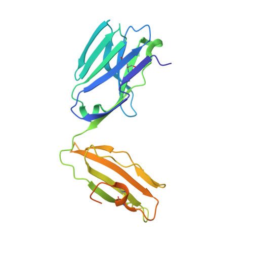
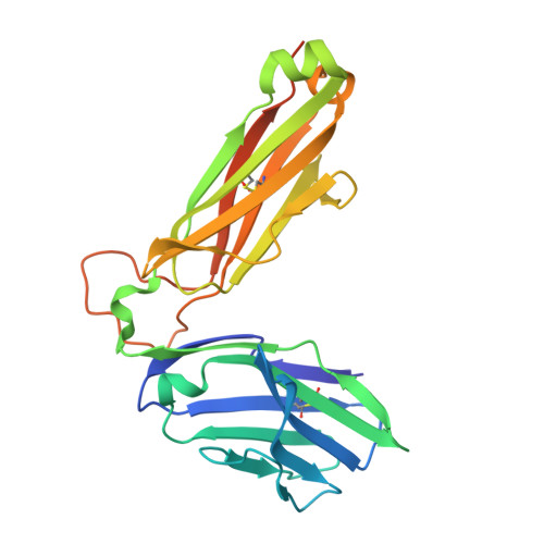
Crystal Structures of Mouse CD1d-iGb3 Complex and its Cognate Valpha14 T Cell Receptor Suggest a Model for Dual Recognition of Foreign and Self Glycolipids.

Zajonc, D.M.Savage, P.B.Bendelac, A.Wilson, I.A.Teyton, L.

(2008) J Mol Biol 377: 1104-1116

  • DOI: https://doi.org/10.1016/j.jmb.2008.01.061
  • Primary Citation of Related Structures:  
    2Q7Y, 2Q86

  • PubMed Abstract: 

    The semi-invariant Valpha14Jalpha18 T cell receptor (TCR) is expressed by regulatory NKT cells and has the unique ability to recognize chemically diverse ligands presented by CD1d. The crystal structure of CD1d complexed to a natural, endogenous ligand, isoglobotrihexosylceramide (iGb3), illustrates the extent of this diversity when compared to the binding of potent, exogenous ligands, such as alpha-galactosylceramide (alpha-GalCer). A single mode of recognition for these two classes of ligands would then appear problematic for a single T cell receptor. However, the Valpha14 TCR adopts two different conformations in the crystal where, in one configuration, the presence of a larger cavity between the two CDR3 regions could accommodate iGb3 and, in the other, a smaller cavity fits alpha-GalCer more snugly. Alternatively, the extended iGb3 headgroup could be "squashed" upon docking of the TCR and accommodated between the CD1 and TCR surfaces. Thus, the same TCR may adopt alternative modes of recognition for these foreign and self-ligands for NKT cell activation.


  • Organizational Affiliation

    Department of Molecular Biology, The Scripps Research Institute, 10550 North Torrey Pines Rd., La Jolla, CA 92037, USA.


Macromolecules
Find similar proteins by:  (by identity cutoff)  |  3D Structure
Entity ID: 1
MoleculeChains Sequence LengthOrganismDetailsImage
Valpha14 TCR
A, C
229Mus musculusMutation(s): 0 
Gene Names: Valpha14
UniProt
Find proteins for P01849 (Mus musculus)
Explore P01849 
Go to UniProtKB:  P01849
Entity Groups  
Sequence Clusters30% Identity50% Identity70% Identity90% Identity95% Identity100% Identity
UniProt GroupP01849
Sequence Annotations
Expand
  • Reference Sequence
Find similar proteins by:  (by identity cutoff)  |  3D Structure
Entity ID: 2
MoleculeChains Sequence LengthOrganismDetailsImage
Vbeta8.2
B, D
254Mus musculusMutation(s): 0 
Gene Names: Vbeta8.2
UniProt
Find proteins for P01851 (Mus musculus)
Explore P01851 
Go to UniProtKB:  P01851
Entity Groups  
Sequence Clusters30% Identity50% Identity70% Identity90% Identity95% Identity100% Identity
UniProt GroupP01851
Sequence Annotations
Expand
  • Reference Sequence
Oligosaccharides

Help

Entity ID: 3
MoleculeChains Length2D Diagram Glycosylation3D Interactions
alpha-D-mannopyranose-(1-6)-beta-D-mannopyranose-(1-4)-2-acetamido-2-deoxy-beta-D-glucopyranose-(1-4)-[alpha-L-fucopyranose-(1-6)]2-acetamido-2-deoxy-beta-D-glucopyranose
E
5N-Glycosylation
Glycosylation Resources
GlyTouCan:  G00395TQ
GlyCosmos:  G00395TQ
GlyGen:  G00395TQ
Entity ID: 4
MoleculeChains Length2D Diagram Glycosylation3D Interactions
beta-D-mannopyranose-(1-4)-2-acetamido-2-deoxy-beta-D-glucopyranose-(1-4)-[alpha-L-fucopyranose-(1-6)]2-acetamido-2-deoxy-beta-D-glucopyranose
F
4N-Glycosylation
Glycosylation Resources
GlyTouCan:  G32152BH
GlyCosmos:  G32152BH
GlyGen:  G32152BH
Small Molecules
Ligands 1 Unique
IDChains Name / Formula / InChI Key2D Diagram3D Interactions
NAG
Query on NAG

Download Ideal Coordinates CCD File 
G [auth B]2-acetamido-2-deoxy-beta-D-glucopyranose
C8 H15 N O6
OVRNDRQMDRJTHS-FMDGEEDCSA-N
Experimental Data & Validation

Experimental Data

  • Method: X-RAY DIFFRACTION
  • Resolution: 1.85 Å
  • R-Value Free: 0.223 
  • R-Value Work: 0.179 
  • R-Value Observed: 0.180 
  • Space Group: P 1 21 1
Unit Cell:
Length ( Å )Angle ( ˚ )
a = 65.293α = 90
b = 150.518β = 117.04
c = 65.814γ = 90
Software Package:
Software NamePurpose
DENZOdata reduction
SCALEPACKdata scaling
REFMACrefinement
PDB_EXTRACTdata extraction
HKL-2000data collection
HKL-2000data reduction
MOLREPphasing

Structure Validation

View Full Validation Report


Ligand Structure Quality Assessment 


Entry History 

Deposition Data

  • Released Date: 2008-04-01 
  • Deposition Author(s): Zajonc, D.M.

Revision History  (Full details and data files)

  • Version 1.0: 2008-04-01
    Type: Initial release
  • Version 1.1: 2011-07-13
    Changes: Non-polymer description, Version format compliance
  • Version 1.2: 2017-10-18
    Changes: Refinement description
  • Version 2.0: 2020-07-29
    Type: Remediation
    Reason: Carbohydrate remediation
    Changes: Atomic model, Data collection, Derived calculations, Structure summary
  • Version 2.1: 2023-08-30
    Changes: Data collection, Database references, Refinement description, Structure summary