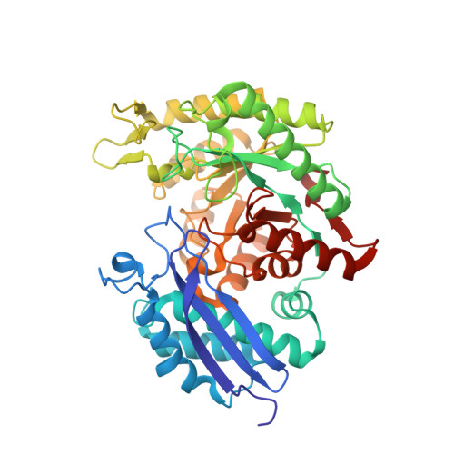
Structural flexibility in Trypanosoma brucei enolase revealed by X-ray crystallography and molecular dynamics.
Navarro, M.V., Gomes Dias, S.M., Mello, L.V., da Silva Giotto, M.T., Gavalda, S., Blonski, C., Garratt, R.C., Rigden, D.J.(2007) FEBS J 274: 5077-5089
- PubMed: 17822439
- DOI: https://doi.org/10.1111/j.1742-4658.2007.06027.x
- Primary Citation of Related Structures:
2PTW, 2PTX, 2PTY, 2PTZ, 2PU0, 2PU1 - PubMed Abstract:
Enolase is a validated drug target in Trypanosoma brucei. To better characterize its properties and guide drug design efforts, we have determined six new crystal structures of the enzyme, in various ligation states and conformations, and have carried out complementary molecular dynamics simulations. The results show a striking structural diversity of loops near the catalytic site, for which variation can be interpreted as distinct modes of conformational variability that are explored during the molecular dynamics simulations. Our results show that sulfate may, unexpectedly, induce full closure of catalytic site loops whereas, conversely, binding of inhibitor phosphonoacetohydroxamate may leave open a tunnel from the catalytic site to protein surface offering possibilities for drug development. We also present the first complex of enolase with a novel inhibitor 2-fluoro-2-phosphonoacetohydroxamate. The molecular dynamics results further encourage efforts to design irreversible species-specific inhibitors: they reveal that a parasite enzyme-specific lysine may approach the catalytic site more closely than crystal structures suggest and also cast light on the issue of accessibility of parasite enzyme-specific cysteines to chemically modifying reagents. One of the new sulfate structures contains a novel metal-binding site IV within the catalytic site cleft.
Organizational Affiliation:
Instituto de Física de São Carlos, Universidade de São Paulo, São Carlos SP, Brazil.