A mixed disulfide bond in bacterial glutathione transferase: functional and evolutionary implications.
Rossjohn, J., Polekhina, G., Feil, S.C., Allocati, N., Masulli, M., De Illio, C., Parker, M.W.(1998) Structure 6: 721-734
- PubMed: 9655824
- DOI: https://doi.org/10.1016/s0969-2126(98)00074-4
- Primary Citation of Related Structures:
1PMT, 2PMT - PubMed Abstract:
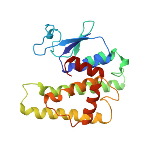
Glutathione S-transferases (GSTs) are a multifunctional group of enzymes, widely distributed in aerobic organisms, that have a critical role in the cellular detoxification process. Unlike their mammalian counterparts, bacterial GSTs often catalyze quite specific reactions, suggesting that their roles in bacteria might be different. The GST from Proteus mirabilis (PmGST B1-1) is known to bind certain antibiotics tightly and reduce the antimicrobial activity of beta-lactam drugs. Hence, bacterial GSTs may play a part in bacterial resistance towards antibiotics and are the subject of intense interest. Here we present the structure of a bacterial GST, PmGST B1-1, which has been determined from two different crystal forms. The enzyme adopts the canonical GST fold although it shares less than 20% sequence identity with GSTs from higher organisms. The most surprising aspect of the structure is the observation that the substrate, glutathione, is covalently bound to Cys 10 of the enzyme. In addition, the highly structurally conserved N-terminal domain is found to have an additional beta strand. The crystal structure of PmGST B1-1 has highlighted the importance of a cysteine residue in the catalytic cycle. Sequence analyses suggest that a number of other GSTs share this property, leading us to propose a new class of GSTs - the beta class. The data suggest that the in vivo role of the beta class GSTs could be as metabolic or redox enzymes rather than conjugating enzymes. Compelling evidence is presented that the theta class of GSTs evolved from an ancestral member of the thioredoxin superfamily.
Organizational Affiliation:
The Ian Potter Foundation Protein Crystallography Laboratory St Vincent's Institute of Medical Research 41 Victoria Parade, Fitzroy, Victoria, 3065, Australia.