2P4M

High pH structure of Rtms5 H146S variant


Experimental Data Snapshot

  • Method: X-RAY DIFFRACTION
  • Resolution: 1.80 Å
  • R-Value Free: 0.249 
  • R-Value Work: 0.226 
  • R-Value Observed: 0.227 

wwPDB Validation   3D Report Full Report


This is version 2.0 of the entry. See complete history


Literature

A structural basis for the pH-dependent increase in fluorescence efficiency of chromoproteins

Battad, J.M.Wilmann, P.G.Olsen, S.Byres, E.Smith, S.C.Dove, S.G.Turcic, K.N.Devenish, R.J.Rossjohn, J.Prescott, M.

(2007) J Mol Biol 368: 998-1010

  • DOI: https://doi.org/10.1016/j.jmb.2007.02.007
  • Primary Citation of Related Structures:  
    2P4M

  • PubMed Abstract: 

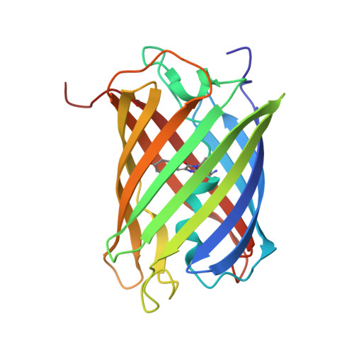
    Within the fluorescent protein and chromoprotein family, the phenomenon of photoswitching is both intriguing and biotechnologically useful. Illumination of particular chromoproteins with intense light results in dramatic increases in fluorescence efficiency (termed kindling) and involves cis-trans isomerization of the chromophore. Here we report that chromophore isomerization can also be driven via alteration in pH. Specifically, we demonstrate that a number of naturally occurring chromoproteins, and their engineered variants, undergo a dramatic 20-100-fold increase in fluorescence efficiency at alkaline pH (>pH9.0). We have determined to 1.8 A resolution the structure of one such chromoprotein, Rtms5(H146S), in its highly far-red fluorescent form (Phi(F), 0.11 at pH 10.7) and compared it to the structure of the non-fluorescent form (Phi(F), 0.002 at pH 8.0). At high pH, the cyclic tri-peptide chromophore was observed to be mobile and distributed between a trans non-coplanar and a cis coplanar conformation, whereas at the lower pH, only a trans non-coplanar chromophore was observed. Calculation of pK(a) values suggested that titration of the side-chain of the conserved Glu215 close to the chromophore is involved in promoting the cis-coplanar conformation. Collectively, our data establish that isomerization to form a coplanar chromophore is a basis of the increased fluorescence efficiency at high pH. The phenomenon of pH-induced fluorescence gain has similarities with photoswitching, thereby providing a model to study the mechanism of kindling.


  • Organizational Affiliation

    Department of Biochemistry and Molecular Biology, School of Biomedical Sciences, Monash University, Clayton Campus, Victoria 3800, Australia.


Macromolecules
Find similar proteins by:  (by identity cutoff)  |  3D Structure
Entity ID: 1
MoleculeChains Sequence LengthOrganismDetailsImage
GFP-like non-fluorescent chromoprotein
A, B, C, D, E
A, B, C, D, E, F, G, H
219Montipora efflorescensMutation(s): 2 
Gene Names: Rtms5
UniProt
Find proteins for P83690 (Montipora efflorescens)
Explore P83690 
Go to UniProtKB:  P83690
Entity Groups  
Sequence Clusters30% Identity50% Identity70% Identity90% Identity95% Identity100% Identity
UniProt GroupP83690
Sequence Annotations
Expand
  • Reference Sequence
Small Molecules
Ligands 1 Unique
IDChains Name / Formula / InChI Key2D Diagram3D Interactions
IOD
Query on IOD

Download Ideal Coordinates CCD File 
AA [auth F]
BA [auth G]
CA [auth G]
DA [auth G]
EA [auth H]
AA [auth F],
BA [auth G],
CA [auth G],
DA [auth G],
EA [auth H],
FA [auth H],
I [auth A],
J [auth A],
K [auth A],
L [auth A],
M [auth B],
N [auth B],
O [auth B],
P [auth B],
Q [auth C],
R [auth C],
S [auth C],
T [auth D],
U [auth D],
V [auth D],
W [auth D],
X [auth E],
Y [auth E],
Z [auth F]
IODIDE ION
I
XMBWDFGMSWQBCA-UHFFFAOYSA-M
Modified Residues  1 Unique
IDChains TypeFormula2D DiagramParent
CRQ
Query on CRQ
A, B, C, D, E
A, B, C, D, E, F, G, H
L-PEPTIDE LINKINGC16 H16 N4 O5GLN, TYR, GLY
Experimental Data & Validation

Experimental Data

  • Method: X-RAY DIFFRACTION
  • Resolution: 1.80 Å
  • R-Value Free: 0.249 
  • R-Value Work: 0.226 
  • R-Value Observed: 0.227 
  • Space Group: C 2 2 21
Unit Cell:
Length ( Å )Angle ( ˚ )
a = 153.75α = 90
b = 186.319β = 90
c = 185.564γ = 90
Software Package:
Software NamePurpose
REFMACrefinement
PDB_EXTRACTdata extraction

Structure Validation

View Full Validation Report


Entry History 

Deposition Data

Revision History  (Full details and data files)

  • Version 1.0: 2007-04-03
    Type: Initial release
  • Version 1.1: 2008-05-01
    Changes: Version format compliance
  • Version 1.2: 2011-07-13
    Changes: Advisory, Version format compliance
  • Version 1.3: 2021-11-10
    Changes: Database references, Derived calculations
  • Version 1.4: 2023-10-25
    Changes: Data collection, Refinement description
  • Version 2.0: 2023-11-15
    Changes: Advisory, Atomic model, Data collection, Derived calculations