Structural analysis of Canavalia maritima and Canavalia gladiata lectins complexed with different dimannosides: new insights into the understanding of the structure-biological activity relationship in legume lectins.
Bezerra, G.A., Oliveira, T.M., Moreno, F.B.M.B., de Souza, E.P., da Rocha, B.A.M., Benevides, R.G., Delatorre, P., de Azevedo Jr., W.F., Cavada, B.S.(2007) J Struct Biol 160: 168-176
- PubMed: 17881248
- DOI: https://doi.org/10.1016/j.jsb.2007.07.012
- Primary Citation of Related Structures:
2EF6, 2OVU, 2OW4, 2P2K, 2P34, 2P37 - PubMed Abstract:
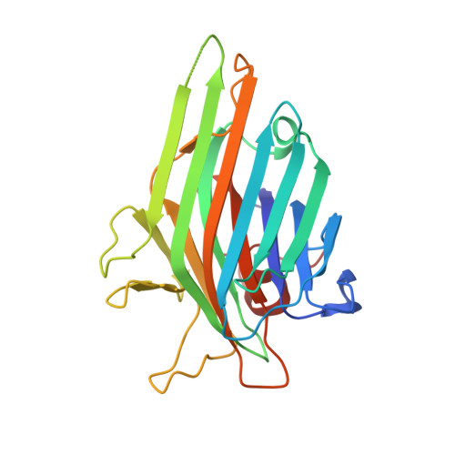
Plant lectins, especially those purified from species of the Leguminosae family, represent the best studied group of carbohydrate-binding proteins. The legume lectins from Diocleinae subtribe are highly similar proteins that present significant differences in the potency/efficacy of their biological activities. The structural studies of the interactions between lectins and sugars may clarify the origin of the distinct biological activities observed in this high similar class of proteins. In this way, this work presents a crystallographic study of the ConM and CGL (agglutinins from Canavalia maritima and Canavalia gladiata, respectively) in the following complexes: ConM/CGL:Man(alpha1-2)Man(alpha1-O)Me, ConM/CGL:Man(alpha1-3)Man(alpha1-O)Me and ConM/CGL:Man(alpha1-4)Man(alpha1-O)Me, which crystallized in different conditions and space group from the native proteins. The structures were solved by molecular replacement, presenting satisfactory values for R(factor) and R(free). Comparisons between ConM, CGL and ConA (Canavalia ensiformis lectin) binding mode with the dimannosides in subject, presented different interactions patterns, which may account for a structural explanation of the distincts biological properties observed in the lectins of Diocleinae subtribe.
Organizational Affiliation:
Departamento de Bioquímica e Biologia Molecular, Universidade Federal do Ceará, Biomol-LAB, Campus do Pici S/N, Fortaleza, Ceará, Brazil.