2OZA

Structure of p38alpha complex


Experimental Data Snapshot

  • Method: X-RAY DIFFRACTION
  • Resolution: 2.70 Å
  • R-Value Free: 0.296 
  • R-Value Work: 0.239 

wwPDB Validation   3D Report Full Report


This is version 1.3 of the entry. See complete history


Literature

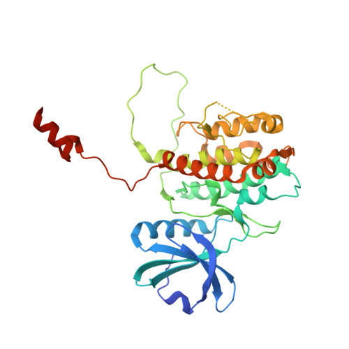
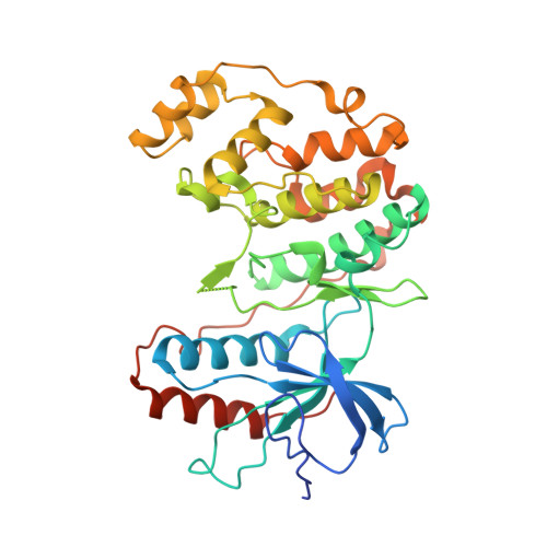
Molecular basis of MAPK-activated protein kinase 2:p38 assembly

White, A.Pargellis, C.A.Studts, J.M.Werneburg, B.G.Farmer II, B.T.

(2007) Proc Natl Acad Sci U S A 104: 6353-6358

  • DOI: https://doi.org/10.1073/pnas.0701679104
  • Primary Citation of Related Structures:  
    2OZA

  • PubMed Abstract: 

    p38 MAPK and MAPK-activated protein kinase 2 (MK2) are key components of signaling pathways leading to many cellular responses, notably the proinflammatory cytokine production. The physical association of p38alpha isoform and MK2 is believed to be physiologically important for this signaling. We report the 2.7-A resolution crystal structure of the unphosphorylated complex between p38alpha and MK2. These protein kinases bind "head-to-head," present their respective active sites on approximately the same side of the heterodimer, and form extensive intermolecular interactions. Among these interactions, the MK2 Ile-366-Ala-390, which includes the bipartite nuclear localization signal, binds to the p38alpha-docking region. This binding supports the involvement of noncatalytic regions to the tight binding of the MK2:p38alpha binary assembly. The MK2 residues 345-365, containing the nuclear export signal, block access to the p38alpha active site. Some regulatory phosphorylation regions of both protein kinases engage in multiple interactions with one another in this complex. This structure gives new insights into the regulation of the protein kinases p38alpha and MK2, aids in the better understanding of their known cellular and biochemical studies, and provides a basis for understanding other regulatory protein-protein interactions involving signal transduction proteins.


  • Organizational Affiliation

    Department of Medicinal Chemistry, Boehringer Ingelheim Pharmaceuticals, Inc., Ridgefield, CT 06877, USA. awhite@rdg.hoehringer-ingelheim.com


Macromolecules
Find similar proteins by:  (by identity cutoff)  |  3D Structure
Entity ID: 1
MoleculeChains Sequence LengthOrganismDetailsImage
MAP kinase-activated protein kinase 2356Homo sapiensMutation(s): 0 
Gene Names: MAPKAPK2
EC: 2.7.11.1
UniProt & NIH Common Fund Data Resources
Find proteins for P49137 (Homo sapiens)
Explore P49137 
Go to UniProtKB:  P49137
PHAROS:  P49137
GTEx:  ENSG00000162889 
Entity Groups  
Sequence Clusters30% Identity50% Identity70% Identity90% Identity95% Identity100% Identity
UniProt GroupP49137
Sequence Annotations
Expand
  • Reference Sequence
Find similar proteins by:  (by identity cutoff)  |  3D Structure
Entity ID: 2
MoleculeChains Sequence LengthOrganismDetailsImage
Mitogen-activated protein kinase 14366Mus musculusMutation(s): 0 
Gene Names: Mapk14Crk1Csbp1Csbp2
EC: 2.7.11.24
UniProt
Find proteins for P47811 (Mus musculus)
Explore P47811 
Go to UniProtKB:  P47811
Entity Groups  
Sequence Clusters30% Identity50% Identity70% Identity90% Identity95% Identity100% Identity
UniProt GroupP47811
Sequence Annotations
Expand
  • Reference Sequence
Experimental Data & Validation

Experimental Data

  • Method: X-RAY DIFFRACTION
  • Resolution: 2.70 Å
  • R-Value Free: 0.296 
  • R-Value Work: 0.239 
  • Space Group: P 41 21 2
Unit Cell:
Length ( Å )Angle ( ˚ )
a = 83.31α = 90
b = 83.31β = 90
c = 231.49γ = 90
Software Package:
Software NamePurpose
d*TREKdata processing
CNSrefinement
PDB_EXTRACTdata extraction
SGX-CATdata collection
d*TREKdata reduction
d*TREKdata scaling
AMoREphasing
CNXrefinement

Structure Validation

View Full Validation Report


Entry History 

Deposition Data

Revision History  (Full details and data files)

  • Version 1.0: 2007-04-03
    Type: Initial release
  • Version 1.1: 2008-05-01
    Changes: Version format compliance
  • Version 1.2: 2011-07-13
    Changes: Version format compliance
  • Version 1.3: 2024-02-21
    Changes: Data collection, Database references