


The crystal structure of recombinant Rhodovulum sulfidophilum SoxAX confirms cysteine persulfide coordination to the catalytic heme
Kihlken, M.A., Berks, B.C., Hemmings, A.M.To be published.
Experimental Data Snapshot
Entity ID: 1 | |||||
|---|---|---|---|---|---|
| Molecule | Chains | Sequence Length | Organism | Details | Image |
| Diheme cytochrome c | 261 | Rhodovulum sulfidophilum | Mutation(s): 1 Gene Names: soxA | 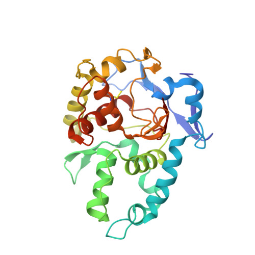 | |
UniProt | |||||
Find proteins for Q939U1 (Rhodovulum sulfidophilum) Explore Q939U1 Go to UniProtKB: Q939U1 | |||||
Entity Groups | |||||
| Sequence Clusters | 30% Identity50% Identity70% Identity90% Identity95% Identity100% Identity | ||||
| UniProt Group | Q939U1 | ||||
Sequence AnnotationsExpand | |||||
| |||||
Entity ID: 2 | |||||
|---|---|---|---|---|---|
| Molecule | Chains | Sequence Length | Organism | Details | Image |
| Cytochrome c | 138 | Rhodovulum sulfidophilum | Mutation(s): 0 Gene Names: soxX | 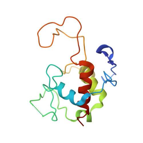 | |
UniProt | |||||
Find proteins for Q939U4 (Rhodovulum sulfidophilum) Explore Q939U4 Go to UniProtKB: Q939U4 | |||||
Entity Groups | |||||
| Sequence Clusters | 30% Identity50% Identity70% Identity90% Identity95% Identity100% Identity | ||||
| UniProt Group | Q939U4 | ||||
Sequence AnnotationsExpand | |||||
| |||||
| Ligands 1 Unique | |||||
|---|---|---|---|---|---|
| ID | Chains | Name / Formula / InChI Key | 2D Diagram | 3D Interactions | |
| HEC Query on HEC | I [auth A] J [auth A] K [auth B] L [auth C] M [auth C] | HEME C C34 H34 Fe N4 O4 HXQIYSLZKNYNMH-LJNAALQVSA-N |  | ||
| Modified Residues 1 Unique | |||||
|---|---|---|---|---|---|
| ID | Chains | Type | Formula | 2D Diagram | Parent |
| CSS Query on CSS | A, C, E, G | L-PEPTIDE LINKING | C3 H7 N O2 S2 |  | CYS |
| Length ( Å ) | Angle ( ˚ ) |
|---|---|
| a = 80.975 | α = 90 |
| b = 102.949 | β = 110.22 |
| c = 115.706 | γ = 90 |
| Software Name | Purpose |
|---|---|
| SCALA | data scaling |
| REFMAC | refinement |
| PDB_EXTRACT | data extraction |
| DNA | data collection |
| MOSFLM | data reduction |
| MOLREP | phasing |
RCSB PDB (citation) is hosted by
RCSB PDB is a member of the

