2ORX

Structural Basis for Ligand Binding and Heparin Mediated Activation of Neuropilin


Experimental Data Snapshot

  • Method: X-RAY DIFFRACTION
  • Resolution: 2.40 Å
  • R-Value Free: 0.252 
  • R-Value Work: 0.201 
  • R-Value Observed: 0.204 

wwPDB Validation   3D Report Full Report


This is version 1.3 of the entry. See complete history


Literature

Structural basis for ligand and heparin binding to neuropilin B domains

Vander Kooi, C.W.Jusino, M.A.Perman, B.Neau, D.B.Bellamy, H.D.Leahy, D.J.

(2007) Proc Natl Acad Sci U S A 104: 6152-6157

  • DOI: https://doi.org/10.1073/pnas.0700043104
  • Primary Citation of Related Structures:  
    2ORX, 2ORZ

  • PubMed Abstract: 

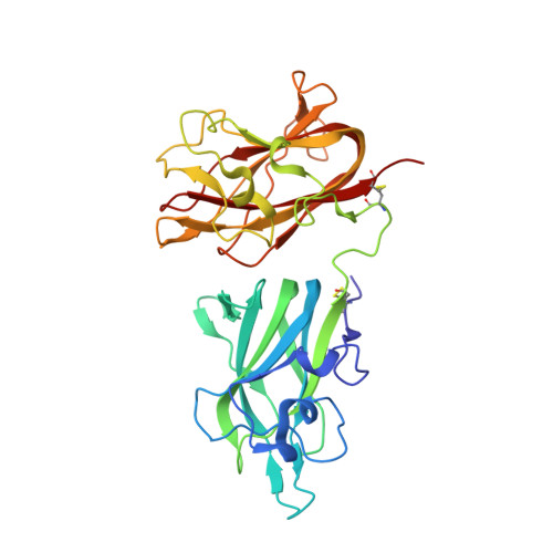
    Neuropilin (Nrp) is a cell surface receptor with essential roles in angiogenesis and axon guidance. Interactions between Nrp and the positively charged C termini of its ligands, VEGF and semaphorin, are mediated by Nrp domains b1 and b2, which share homology to coagulation factor domains. We report here the crystal structure of the tandem b1 and b2 domains of Nrp-1 (N1b1b2) and show that they form a single structural unit. Cocrystallization of N1b1b2 with Tuftsin, a peptide mimic of the VEGF C terminus, reveals the site of interaction with the basic tail of VEGF on the b1 domain. We also show that heparin promotes N1b1b2 dimerization and map the heparin binding site on N1b1b2. These results provide a detailed picture of interactions at the core of the Nrp signaling complex and establish a molecular basis for the synergistic effects of heparin on Nrp-mediated signaling.


  • Organizational Affiliation

    Department of Biophysics and Biophysical Chemistry, Johns Hopkins University School of Medicine, Baltimore, MD 21205, USA.


Macromolecules
Find similar proteins by:  (by identity cutoff)  |  3D Structure
Entity ID: 1
MoleculeChains Sequence LengthOrganismDetailsImage
Neuropilin-1314Rattus norvegicusMutation(s): 0 
Gene Names: Nrp1
UniProt
Find proteins for Q9QWJ9 (Rattus norvegicus)
Explore Q9QWJ9 
Go to UniProtKB:  Q9QWJ9
Entity Groups  
Sequence Clusters30% Identity50% Identity70% Identity90% Identity95% Identity100% Identity
UniProt GroupQ9QWJ9
Sequence Annotations
Expand
  • Reference Sequence
Experimental Data & Validation

Experimental Data

  • Method: X-RAY DIFFRACTION
  • Resolution: 2.40 Å
  • R-Value Free: 0.252 
  • R-Value Work: 0.201 
  • R-Value Observed: 0.204 
  • Space Group: P 21 21 21
Unit Cell:
Length ( Å )Angle ( ˚ )
a = 38.094α = 90
b = 92.221β = 90
c = 120.115γ = 90
Software Package:
Software NamePurpose
REFMACrefinement
HKL-3000data collection
HKL-3000data reduction
HKL-3000data scaling
MOLREPphasing

Structure Validation

View Full Validation Report


Entry History 

Deposition Data

Revision History  (Full details and data files)

  • Version 1.0: 2007-04-03
    Type: Initial release
  • Version 1.1: 2008-04-21
    Changes: Version format compliance
  • Version 1.2: 2011-07-13
    Changes: Advisory, Version format compliance
  • Version 1.3: 2023-08-30
    Changes: Data collection, Database references, Refinement description