2ON9

Structure of an amyloid forming peptide VQIVYK from the repeat region of Tau


Experimental Data Snapshot

  • Method: X-RAY DIFFRACTION
  • Resolution: 1.51 Å
  • R-Value Free: 0.225 
  • R-Value Work: 0.206 
  • R-Value Observed: 0.207 

wwPDB Validation   3D Report Full Report


This is version 1.5 of the entry. See complete history


Literature

Atomic structures of amyloid cross-beta spines reveal varied steric zippers.

Sawaya, M.R.Sambashivan, S.Nelson, R.Ivanova, M.I.Sievers, S.A.Apostol, M.I.Thompson, M.J.Balbirnie, M.Wiltzius, J.J.McFarlane, H.T.Madsen, A.O.Riekel, C.Eisenberg, D.

(2007) Nature 447: 453-457

  • DOI: https://doi.org/10.1038/nature05695
  • Primary Citation of Related Structures:  
    2OKZ, 2OL9, 2OLX, 2OMM, 2OMP, 2OMQ, 2ON9, 2ONA, 2ONV, 2ONW, 2ONX

  • PubMed Abstract: 

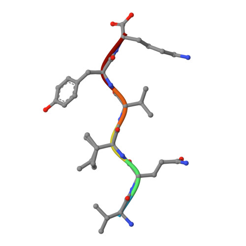
    Amyloid fibrils formed from different proteins, each associated with a particular disease, contain a common cross-beta spine. The atomic architecture of a spine, from the fibril-forming segment GNNQQNY of the yeast prion protein Sup35, was recently revealed by X-ray microcrystallography. It is a pair of beta-sheets, with the facing side chains of the two sheets interdigitated in a dry 'steric zipper'. Here we report some 30 other segments from fibril-forming proteins that form amyloid-like fibrils, microcrystals, or usually both. These include segments from the Alzheimer's amyloid-beta and tau proteins, the PrP prion protein, insulin, islet amyloid polypeptide (IAPP), lysozyme, myoglobin, alpha-synuclein and beta(2)-microglobulin, suggesting that common structural features are shared by amyloid diseases at the molecular level. Structures of 13 of these microcrystals all reveal steric zippers, but with variations that expand the range of atomic architectures for amyloid-like fibrils and offer an atomic-level hypothesis for the basis of prion strains.


  • Organizational Affiliation

    Howard Hughes Medical Institute, UCLA-DOE Institute of Genomics and Proteomics, Los Angeles, California 90095-1570, USA.


Macromolecules

Find similar proteins by:  Sequence   |   3D Structure  

Entity ID: 1
MoleculeChains Sequence LengthOrganismDetailsImage
VQIVYK peptide corresponding to residues 306-311 in the tau protein
A, B
6N/AMutation(s): 0 
UniProt & NIH Common Fund Data Resources
Find proteins for P10636 (Homo sapiens)
Explore P10636 
Go to UniProtKB:  P10636
PHAROS:  P10636
GTEx:  ENSG00000186868 
Entity Groups  
UniProt GroupP10636
Sequence Annotations
Expand
  • Reference Sequence
Experimental Data & Validation

Experimental Data

  • Method: X-RAY DIFFRACTION
  • Resolution: 1.51 Å
  • R-Value Free: 0.225 
  • R-Value Work: 0.206 
  • R-Value Observed: 0.207 
  • Space Group: P 1 21 1
Unit Cell:
Length ( Å )Angle ( ˚ )
a = 4.863α = 90
b = 61.926β = 98.11
c = 15.413γ = 90
Software Package:
Software NamePurpose
DENZOdata reduction
SCALEPACKdata scaling
PHASERphasing
REFMACrefinement
PDB_EXTRACTdata extraction

Structure Validation

View Full Validation Report


Entry History 

Deposition Data

Revision History  (Full details and data files)

  • Version 1.0: 2007-01-30
    Type: Initial release
  • Version 1.1: 2008-05-01
    Changes: Version format compliance
  • Version 1.2: 2011-07-13
    Changes: Version format compliance
  • Version 1.3: 2017-10-18
    Changes: Refinement description
  • Version 1.4: 2023-12-27
    Changes: Data collection, Database references
  • Version 1.5: 2024-04-03
    Changes: Refinement description