2OL3

crystal structure of BM3.3 ScFV TCR in complex with PBM8-H-2KBM8 MHC class I molecule


Experimental Data Snapshot

  • Method: X-RAY DIFFRACTION
  • Resolution: 2.90 Å
  • R-Value Free: 0.284 
  • R-Value Work: 0.224 
  • R-Value Observed: 0.227 

wwPDB Validation   3D Report Full Report


Ligand Structure Quality Assessment 


This is version 1.4 of the entry. See complete history


Literature

How much can a T-cell antigen receptor adapt to structurally distinct antigenic peptides?

Mazza, C.Auphan-Anezin, N.Gregoire, C.Guimezanes, A.Kellenberger, C.Roussel, A.Kearney, A.van der Merwe, P.A.Schmitt-Verhulst, A.M.Malissen, B.

(2007) EMBO J 26: 1972-1983

  • DOI: https://doi.org/10.1038/sj.emboj.7601605
  • Primary Citation of Related Structures:  
    2OL3

  • PubMed Abstract: 

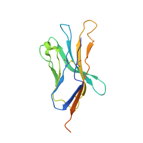
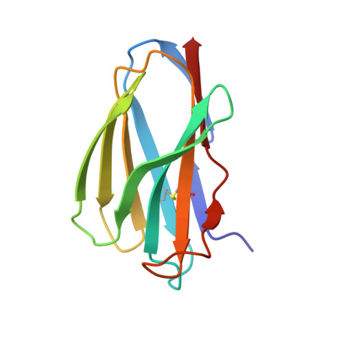
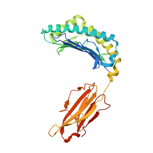
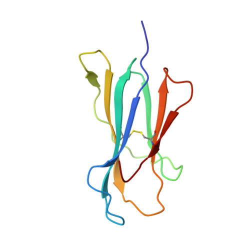
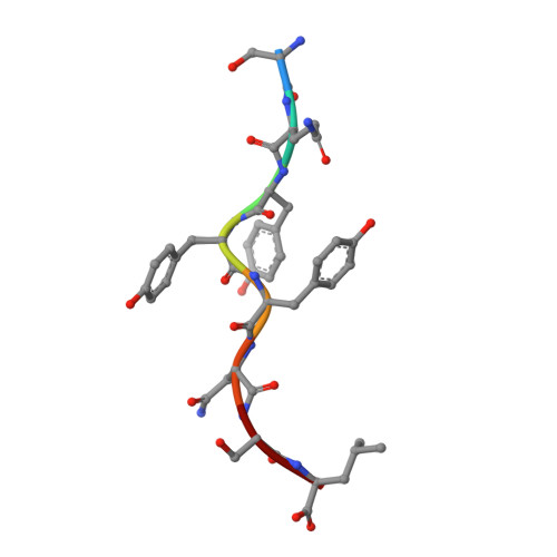
    Binding degeneracy is thought to constitute a fundamental property of the T-cell antigen receptor (TCR), yet its structural basis is poorly understood. We determined the crystal structure of a complex involving the BM3.3 TCR and a peptide (pBM8) bound to the H-2K(bm8) major histocompatibility complex (MHC) molecule, and compared it with the structures of the BM3.3 TCR bound to H-2K(b) molecules loaded with two peptides that had a minimal level of primary sequence identity with pBM8. Our findings provide a refined structural view of the basis of BM3.3 TCR cross-reactivity and a structural explanation for the long-standing paradox that a TCR antigen-binding site can be both specific and degenerate. We also measured the thermodynamic features and biological penalties that incurred during cross-recognition. Our data illustrate the difficulty for a given TCR in adapting to distinct peptide-MHC surfaces while still maintaining affinities that result in functional in vivo responses. Therefore, when induction of protective effector T cells is used as the ultimate criteria for adaptive immunity, TCRs are probably much less degenerate than initially assumed.


  • Organizational Affiliation

    Centre d'Immunologie de Marseille-Luminy, Université de la Méditerrannée, 13288 Marseille Cedex 09, France.


Macromolecules
Find similar proteins by:  (by identity cutoff)  |  3D Structure
Entity ID: 1
MoleculeChains Sequence LengthOrganismDetailsImage
BM3.3 T-CELL RECEPTOR ALPHA-CHAIN142Mus musculusMutation(s): 0 
UniProt
Find proteins for Q5R1F1 (Mus musculus)
Explore Q5R1F1 
Go to UniProtKB:  Q5R1F1
Entity Groups  
Sequence Clusters30% Identity50% Identity70% Identity90% Identity95% Identity100% Identity
UniProt GroupQ5R1F1
Sequence Annotations
Expand
  • Reference Sequence
Find similar proteins by:  (by identity cutoff)  |  3D Structure
Entity ID: 2
MoleculeChains Sequence LengthOrganismDetailsImage
BM3.3 T-CELL RECEPTOR BETA-CHAIN113Mus musculusMutation(s): 0 
UniProt
Find proteins for P04214 (Mus musculus)
Explore P04214 
Go to UniProtKB:  P04214
Entity Groups  
Sequence Clusters30% Identity50% Identity70% Identity90% Identity95% Identity100% Identity
UniProt GroupP04214
Sequence Annotations
Expand
  • Reference Sequence
Find similar proteins by:  (by identity cutoff)  |  3D Structure
Entity ID: 3
MoleculeChains Sequence LengthOrganismDetailsImage
ALLOGENEIC H-2KBM8 MHC CLASS I MOLECULEC [auth H]279Mus musculusMutation(s): 0 
UniProt
Find proteins for P01901 (Mus musculus)
Explore P01901 
Go to UniProtKB:  P01901
Entity Groups  
Sequence Clusters30% Identity50% Identity70% Identity90% Identity95% Identity100% Identity
UniProt GroupP01901
Sequence Annotations
Expand
  • Reference Sequence
Find similar proteins by:  (by identity cutoff)  |  3D Structure
Entity ID: 4
MoleculeChains Sequence LengthOrganismDetailsImage
Beta-2-microglobulinD [auth L]100Mus musculusMutation(s): 0 
Gene Names: B2m
UniProt & NIH Common Fund Data Resources
Find proteins for P01887 (Mus musculus)
Explore P01887 
Go to UniProtKB:  P01887
IMPC:  MGI:88127
Entity Groups  
Sequence Clusters30% Identity50% Identity70% Identity90% Identity95% Identity100% Identity
UniProt GroupP01887
Sequence Annotations
Expand
  • Reference Sequence

Find similar proteins by:  Sequence   |   3D Structure  

Entity ID: 5
MoleculeChains Sequence LengthOrganismDetailsImage
NATURALLY PROCESSED OCTAPEPTIDE PBM8E [auth P]8N/AMutation(s): 0 
UniProt
Find proteins for Q91YE7 (Mus musculus)
Explore Q91YE7 
Go to UniProtKB:  Q91YE7
Entity Groups  
UniProt GroupQ91YE7
Sequence Annotations
Expand
  • Reference Sequence
Small Molecules
Ligands 1 Unique
IDChains Name / Formula / InChI Key2D Diagram3D Interactions
NAG
Query on NAG

Download Ideal Coordinates CCD File 
F [auth A]2-acetamido-2-deoxy-beta-D-glucopyranose
C8 H15 N O6
OVRNDRQMDRJTHS-FMDGEEDCSA-N
Experimental Data & Validation

Experimental Data

  • Method: X-RAY DIFFRACTION
  • Resolution: 2.90 Å
  • R-Value Free: 0.284 
  • R-Value Work: 0.224 
  • R-Value Observed: 0.227 
  • Space Group: P 43 21 2
Unit Cell:
Length ( Å )Angle ( ˚ )
a = 102.523α = 90
b = 102.523β = 90
c = 198.381γ = 90
Software Package:
Software NamePurpose
REFMACrefinement
MOSFLMdata reduction
CCP4data scaling
AMoREphasing

Structure Validation

View Full Validation Report


Ligand Structure Quality Assessment 


Entry History 

Revision History  (Full details and data files)

  • Version 1.0: 2007-04-03
    Type: Initial release
  • Version 1.1: 2008-05-01
    Changes: Version format compliance
  • Version 1.2: 2011-07-13
    Changes: Advisory, Version format compliance
  • Version 1.3: 2020-07-29
    Type: Remediation
    Reason: Carbohydrate remediation
    Changes: Data collection, Database references, Derived calculations, Structure summary
  • Version 1.4: 2023-08-30
    Changes: Data collection, Database references, Refinement description, Structure summary