2O7L

The open-cap conformation of GlpG


Experimental Data Snapshot

  • Method: X-RAY DIFFRACTION
  • Resolution: 2.50 Å
  • R-Value Free: 0.285 
  • R-Value Work: 0.240 

wwPDB Validation   3D Report Full Report


Ligand Structure Quality Assessment 


This is version 1.4 of the entry. See complete history


Literature

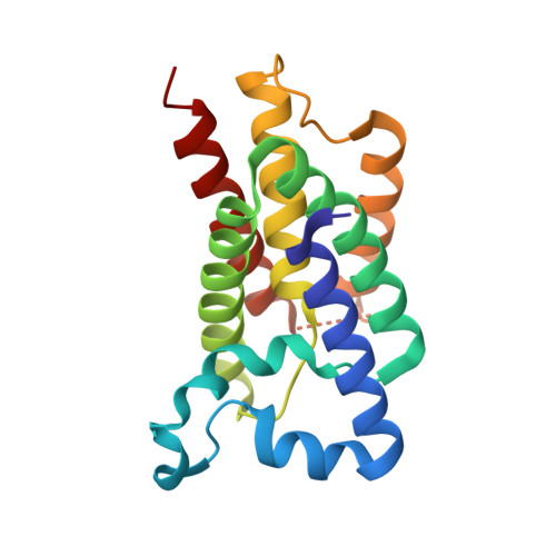
Open-cap conformation of intramembrane protease GlpG.

Wang, Y.Ha, Y.

(2007) Proc Natl Acad Sci U S A 104: 2098-2102

  • DOI: https://doi.org/10.1073/pnas.0611080104
  • Primary Citation of Related Structures:  
    2O7L

  • PubMed Abstract: 

    The active sites of intramembrane proteases are positioned in the lipid bilayer to facilitate peptide bond hydrolysis in the membrane. Previous crystallographic analysis of Escherichia coli GlpG, an intramembrane protease of the rhomboid family, has revealed an internal and hydrophilic active site in an apparently closed conformation. Here we describe the crystal structure of GlpG in a more open conformation, where the capping loop L5 has been lifted, exposing the previously buried and catalytically essential Ser-201 to outside aqueous solution. A water molecule now moves into the putative oxyanion hole that is constituted of a main-chain amide (Ser-201) and two conserved side chains (His-150 and Asn-154). The loop movement also destabilizes a hydrophobic side chain (Phe-245) previously buried between transmembrane helices S2 and S5 and creates a side portal from the lipid to protease active site. These results provide insights into the conformational plasticity of GlpG to accommodate substrate binding and catalysis and into the chirality of the reaction intermediate.


  • Organizational Affiliation

    Department of Pharmacology, Yale University School of Medicine, 333 Cedar Street, New Haven, CT 06520, USA.


Macromolecules
Find similar proteins by:  (by identity cutoff)  |  3D Structure
Entity ID: 1
MoleculeChains Sequence LengthOrganismDetailsImage
Protein glpG180Escherichia coliMutation(s): 0 
Gene Names: glpG
Membrane Entity: Yes 
UniProt
Find proteins for P09391 (Escherichia coli (strain K12))
Explore P09391 
Go to UniProtKB:  P09391
Entity Groups  
Sequence Clusters30% Identity50% Identity70% Identity90% Identity95% Identity100% Identity
UniProt GroupP09391
Sequence Annotations
Expand
  • Reference Sequence
Small Molecules
Ligands 1 Unique
IDChains Name / Formula / InChI Key2D Diagram3D Interactions
BNG
Query on BNG

Download Ideal Coordinates CCD File 
B [auth A]nonyl beta-D-glucopyranoside
C15 H30 O6
QFAPUKLCALRPLH-UXXRCYHCSA-N
Experimental Data & Validation

Experimental Data

  • Method: X-RAY DIFFRACTION
  • Resolution: 2.50 Å
  • R-Value Free: 0.285 
  • R-Value Work: 0.240 
  • Space Group: H 3 2
Unit Cell:
Length ( Å )Angle ( ˚ )
a = 111.4α = 90
b = 111.4β = 90
c = 128.6γ = 120
Software Package:
Software NamePurpose
CNSrefinement
PDB_EXTRACTdata extraction
HKL-2000data collection
HKL-2000data reduction
HKL-2000data scaling
CNSphasing

Structure Validation

View Full Validation Report


Ligand Structure Quality Assessment 


Entry History 

Deposition Data

  • Released Date: 2006-12-26 
  • Deposition Author(s): Ha, Y.

Revision History  (Full details and data files)

  • Version 1.0: 2006-12-26
    Type: Initial release
  • Version 1.1: 2008-05-01
    Changes: Version format compliance
  • Version 1.2: 2011-07-13
    Changes: Version format compliance
  • Version 1.3: 2020-07-29
    Type: Remediation
    Reason: Carbohydrate remediation
    Changes: Data collection, Derived calculations, Structure summary
  • Version 1.4: 2023-08-30
    Changes: Data collection, Database references, Refinement description, Structure summary