2NGR

TRANSITION STATE COMPLEX FOR GTP HYDROLYSIS BY CDC42: COMPARISONS OF THE HIGH RESOLUTION STRUCTURES FOR CDC42 BOUND TO THE ACTIVE AND CATALYTICALLY COMPROMISED FORMS OF THE CDC42-GAP.


Experimental Data Snapshot

  • Method: X-RAY DIFFRACTION
  • Resolution: 1.90 Å
  • R-Value Free: 0.293 
  • R-Value Work: 0.253 
  • R-Value Observed: 0.253 

wwPDB Validation   3D Report Full Report


This is version 1.4 of the entry. See complete history


Literature

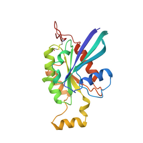
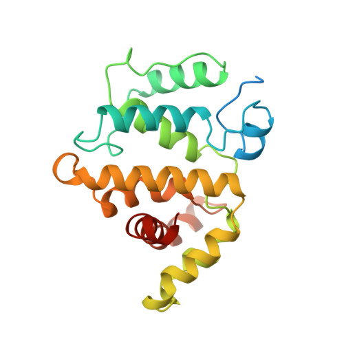
Structures of Cdc42 bound to the active and catalytically compromised forms of Cdc42GAP.

Nassar, N.Hoffman, G.R.Manor, D.Clardy, J.C.Cerione, R.A.

(1998) Nat Struct Biol 5: 1047-1052

  • DOI: https://doi.org/10.1038/4156
  • Primary Citation of Related Structures:  
    1GRN, 2NGR

  • PubMed Abstract: 

    The Rho-related small GTP-binding protein Cdc42 has a low intrinsic GTPase activity that is significantly enhanced by its specific GTPase-activating protein, Cdc42GAP. In this report, we present the tertiary structure for the aluminum fluoride-promoted complex between Cdc42 and a catalytically active domain of Cdc42GAP as well as the complex between Cdc42 and the catalytically compromised Cdc42GAP(R305A) mutant. These structures, which mimic the transition state for the GTP hydrolytic reaction, show the presence of an AIF3 molecule, as was seen for the corresponding Ras-p120RasGAP complex, but in contrast to what has been reported for the Rho-Cdc42GAP complex or for heterotrimeric G protein alpha subunits, where AIF4- was observed. The Cdc42GAP stabilizes both the switch I and switch II domains of Cdc42 and contributes a highly conserved arginine (Arg 305) to the active site. Comparison of the structures for the wild type and mutant Cdc42GAP complexes provides important insights into the GAP-catalyzed GTP hydrolytic reaction.


  • Organizational Affiliation

    Department of Pharmacology, College of Veterinary Medicine, Cornell University, Ithaca, New York 14853, USA.


Macromolecules
Find similar proteins by:  (by identity cutoff)  |  3D Structure
Entity ID: 1
MoleculeChains Sequence LengthOrganismDetailsImage
PROTEIN (GTP BINDING PROTEIN (G25K))191Homo sapiensMutation(s): 0 
UniProt & NIH Common Fund Data Resources
Find proteins for P60953 (Homo sapiens)
Explore P60953 
Go to UniProtKB:  P60953
PHAROS:  P60953
GTEx:  ENSG00000070831 
Entity Groups  
Sequence Clusters30% Identity50% Identity70% Identity90% Identity95% Identity100% Identity
UniProt GroupP60953
Sequence Annotations
Expand
  • Reference Sequence
Find similar proteins by:  (by identity cutoff)  |  3D Structure
Entity ID: 2
MoleculeChains Sequence LengthOrganismDetailsImage
PROTEIN (GTPASE ACTIVATING PROTEIN (RHG))234Homo sapiensMutation(s): 1 
UniProt & NIH Common Fund Data Resources
Find proteins for Q07960 (Homo sapiens)
Explore Q07960 
Go to UniProtKB:  Q07960
PHAROS:  Q07960
GTEx:  ENSG00000175220 
Entity Groups  
Sequence Clusters30% Identity50% Identity70% Identity90% Identity95% Identity100% Identity
UniProt GroupQ07960
Sequence Annotations
Expand
  • Reference Sequence
Small Molecules
Ligands 3 Unique
IDChains Name / Formula / InChI Key2D Diagram3D Interactions
GDP
Query on GDP

Download Ideal Coordinates CCD File 
D [auth A]GUANOSINE-5'-DIPHOSPHATE
C10 H15 N5 O11 P2
QGWNDRXFNXRZMB-UUOKFMHZSA-N
AF3
Query on AF3

Download Ideal Coordinates CCD File 
E [auth A]ALUMINUM FLUORIDE
Al F3
KLZUFWVZNOTSEM-UHFFFAOYSA-K
MG
Query on MG

Download Ideal Coordinates CCD File 
C [auth A]MAGNESIUM ION
Mg
JLVVSXFLKOJNIY-UHFFFAOYSA-N
Experimental Data & Validation

Experimental Data

  • Method: X-RAY DIFFRACTION
  • Resolution: 1.90 Å
  • R-Value Free: 0.293 
  • R-Value Work: 0.253 
  • R-Value Observed: 0.253 
  • Space Group: P 21 21 21
Unit Cell:
Length ( Å )Angle ( ˚ )
a = 52.82α = 90
b = 67.69β = 90
c = 131.06γ = 90
Software Package:
Software NamePurpose
MOSFLMdata reduction
SCALAdata scaling
X-PLORmodel building
X-PLORrefinement
CCP4data scaling
X-PLORphasing

Structure Validation

View Full Validation Report


Entry History 

Deposition Data

Revision History  (Full details and data files)

  • Version 1.0: 1999-01-06
    Type: Initial release
  • Version 1.1: 2008-04-27
    Changes: Version format compliance
  • Version 1.2: 2011-07-13
    Changes: Version format compliance
  • Version 1.3: 2018-03-14
    Changes: Database references
  • Version 1.4: 2023-08-30
    Changes: Data collection, Database references, Derived calculations, Refinement description