2MPA

BACTERICIDAL ANTIBODY AGAINST NEISSERIA MENINGITIDIS


Experimental Data Snapshot

  • Method: X-RAY DIFFRACTION
  • Resolution: 2.60 Å
  • R-Value Free: 0.264 
  • R-Value Work: 0.202 
  • R-Value Observed: 0.202 

wwPDB Validation   3D Report Full Report


This is version 2.0 of the entry. See complete history


Literature

Bactericidal antibody recognition of a PorA epitope of Neisseria meningitidis: crystal structure of a Fab fragment in complex with a fluorescein-conjugated peptide.

van den Elsen, J.M.Herron, J.N.Hoogerhout, P.Poolman, J.T.Boel, E.Logtenberg, T.Wilting, J.Crommelin, D.J.Kroon, J.Gros, P.

(1997) Proteins 29: 113-125

  • DOI: https://doi.org/10.1002/(sici)1097-0134(199709)29:1<113::aid-prot9>3.3.co;2-u
  • Primary Citation of Related Structures:  
    1MPA, 2MPA

  • PubMed Abstract: 

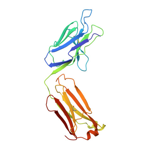
    Class 1 outer membrane protein PorA of Neisseria meningitidis is a vaccine candidate against bacterial meningitis. Antibodies against PorA are able to induce complement-mediated bacterial killing and thereby play an important role in protection against meningococcal disease. Bactericidal antibodies are all directed against variable regions VR1 and VR2 of the PorA sequence, corresponding to loops 1 and 4 of a two-dimensional topology model of the porin with eight extracellular loops. We have determined the crystal structure to 2.6 A resolution of the Fab fragment of bactericidal antibody MN12H2 against meningococcal PorA in complex with a linear fluorescein-conjugated peptide TKDTNNNL derived from the VR2 sequence of sero-subtype P1.7,16 (residues 180-187) from meningococcal strain H44/76. The peptide folds deeply into the binding cavity of the Fab molecule in a type I beta-turn, with the minimal P1.16 epitope DTNNN virtually completely buried. The structure reveals H-bonds and van der Waals interactions with all minimal epitope residues and one essential salt bridge between Asp-182 of the peptide and His-31 of the MN12H2 light chain. The key components of the recognition of PorA epitope P1.16 by bactericidal antibody MN12H2 correspond well with available thermodynamic data from binding studies. Furthermore, they indicate the structural basis of an increased endemic incidence of infection by group B meningococci in England and Wales since 1981 associated with the occurrence of an Neisseria meningitidis escape mutant (strain-MC58). The observed three-dimensional conformation of the peptide provides a rationale for the development of a synthetic peptide vaccine against meningococcal disease.


  • Organizational Affiliation

    Department of Pharmaceutics, Faculty of Pharmacy, Utrecht Institute of Pharmaceutical Sciences, Utrecht University, The Netherlands.


Macromolecules
Find similar proteins by:  (by identity cutoff)  |  3D Structure
Entity ID: 1
MoleculeChains Sequence LengthOrganismDetailsImage
MN12H2 IGG2A-KAPPA, light chainA [auth L]219Mus musculusMutation(s): 0 
UniProt
Find proteins for A2NHM3 (Mus musculus)
Explore A2NHM3 
Go to UniProtKB:  A2NHM3
Entity Groups  
Sequence Clusters30% Identity50% Identity70% Identity90% Identity95% Identity100% Identity
UniProt GroupA2NHM3
Sequence Annotations
Expand
  • Reference Sequence
Find similar proteins by:  (by identity cutoff)  |  3D Structure
Entity ID: 2
MoleculeChains Sequence LengthOrganismDetailsImage
MN12H2 IGG2A-KAPPA,heavy chainB [auth H]225Mus musculusMutation(s): 0 
UniProt
Find proteins for Q569X1 (Mus musculus)
Explore Q569X1 
Go to UniProtKB:  Q569X1
Entity Groups  
Sequence Clusters30% Identity50% Identity70% Identity90% Identity95% Identity100% Identity
UniProt GroupQ569X1
Sequence Annotations
Expand
  • Reference Sequence

Find similar proteins by:  Sequence   |   3D Structure  

Entity ID: 3
MoleculeChains Sequence LengthOrganismDetailsImage
CONJUGATE OF PORA P1.16 PEPTIDE WITH FLUORESCEINC [auth P]9synthetic constructMutation(s): 0 
UniProt
Find proteins for E6MXW0 (Neisseria meningitidis serogroup B / serotype 15 (strain H44/76))
Explore E6MXW0 
Go to UniProtKB:  E6MXW0
Entity Groups  
UniProt GroupE6MXW0
Sequence Annotations
Expand
  • Reference Sequence
Small Molecules
Ligands 1 Unique
IDChains Name / Formula / InChI Key2D Diagram3D Interactions
CD
Query on CD

Download Ideal Coordinates CCD File 
D [auth L]CADMIUM ION
Cd
WLZRMCYVCSSEQC-UHFFFAOYSA-N
Modified Residues  2 Unique
IDChains TypeFormula2D DiagramParent
CYF
Query on CYF
C [auth P]L-PEPTIDE LINKINGC25 H23 N3 O7 SCYS
THC
Query on THC
C [auth P]L-PEPTIDE LINKINGC6 H11 N O4THR
Experimental Data & Validation

Experimental Data

  • Method: X-RAY DIFFRACTION
  • Resolution: 2.60 Å
  • R-Value Free: 0.264 
  • R-Value Work: 0.202 
  • R-Value Observed: 0.202 
  • Space Group: P 1 21 1
Unit Cell:
Length ( Å )Angle ( ˚ )
a = 55.68α = 90
b = 69.1β = 112.01
c = 72.95γ = 90
Software Package:
Software NamePurpose
DENZOdata reduction
SCALEPACKdata scaling
CNSrefinement
CNSphasing

Structure Validation

View Full Validation Report


Entry History 

Deposition Data

Revision History  (Full details and data files)

  • Version 1.0: 1999-06-23
    Type: Initial release
  • Version 1.1: 2007-10-16
    Changes: Version format compliance
  • Version 1.2: 2011-07-13
    Changes: Derived calculations, Version format compliance
  • Version 1.3: 2011-10-05
    Changes: Database references, Derived calculations
  • Version 1.4: 2017-05-31
    Changes: Database references, Source and taxonomy, Structure summary
  • Version 1.5: 2023-08-30
    Changes: Data collection, Database references, Derived calculations, Refinement description
  • Version 2.0: 2023-11-15
    Changes: Atomic model, Data collection