Conopeptide rho-TIA defines a new allosteric site on the extracellular surface of the alpha 1B-adrenoceptor.
Ragnarsson, L., Wang, C.I., Andersson, A., Fajarningsih, D., Monks, T., Brust, A., Rosengren, K.J., Lewis, R.J.(2013) J Biol Chem 288: 1814-1827
- PubMed: 23184947
- DOI: https://doi.org/10.1074/jbc.M112.430785
- Primary Citation of Related Structures:
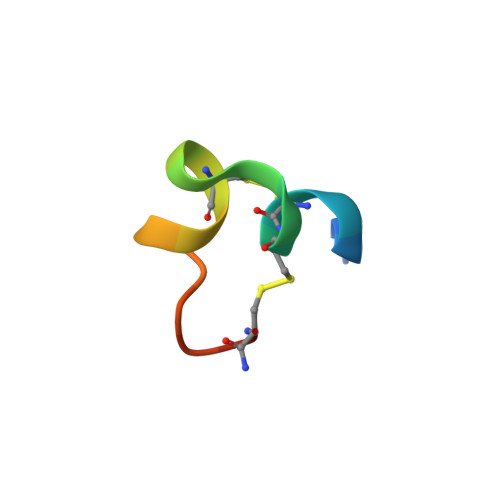
2LR9 - PubMed Abstract:
The G protein-coupled receptor (GPCR) superfamily is an important drug target that includes over 1000 membrane receptors that functionally couple extracellular stimuli to intracellular effectors. Despite the potential of extracellular surface (ECS) residues in GPCRs to interact with subtype-specific allosteric modulators, few ECS pharmacophores for class A receptors have been identified. Using the turkey β(1)-adrenergic receptor crystal structure, we modeled the α(1B)-adrenoceptor (α(1B)-AR) to help identify the allosteric site for ρ-conopeptide TIA, an inverse agonist at this receptor. Combining mutational radioligand binding and inositol 1-phosphate signaling studies, together with molecular docking simulations using a refined NMR structure of ρ-TIA, we identified 14 residues on the ECS of the α(1B)-AR that influenced ρ-TIA binding. Double mutant cycle analysis and docking confirmed that ρ-TIA binding was dominated by a salt bridge and cation-π between Arg-4-ρ-TIA and Asp-327 and Phe-330, respectively, and a T-stacking-π interaction between Trp-3-ρ-TIA and Phe-330. Water-bridging hydrogen bonds between Asn-2-ρ-TIA and Val-197, Trp-3-ρ-TIA and Ser-318, and the positively charged N terminus and Glu-186, were also identified. These interactions reveal that peptide binding to the ECS on transmembrane helix 6 (TMH6) and TMH7 at the base of extracellular loop 3 (ECL3) is sufficient to allosterically inhibit agonist signaling at a GPCR. The ligand-accessible ECS residues identified provide the first view of an allosteric inhibitor pharmacophore for α(1)-adrenoceptors and mechanistic insight and a new set of structural constraints for the design of allosteric antagonists at related GPCRs.
Organizational Affiliation:
Institute for Molecular Bioscience, University of Queensland, Brisbane, Queensland 4072, Australia.