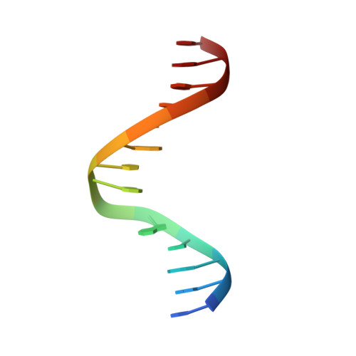
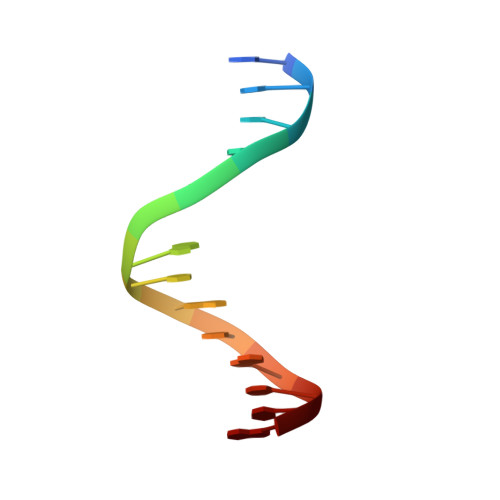
NMR solution structures of clustered abasic site lesions in DNA: structural differences between 3'-staggered (-3) and 5'-staggered (+3) bistranded lesions.
Hazel, R.D., de los Santos, C.(2010) Biochemistry 49: 8978-8987
- PubMed: 20825249
- DOI: https://doi.org/10.1021/bi101021e
- Primary Citation of Related Structures:
2L2U, 2L2V - PubMed Abstract:
Ionizing radiation produces a distinctive pattern of bistranded clustered lesions in DNA. A relatively low number of clustered lesions may be lethal to cells when compared to a larger number of single lesions. Enzyme cleavage experiments suggest that the orientation of bistranded lesions causes differential recognition and removal of these lesions. Like that of a previous study of bistranded abasic site lesion [Hazel, R. D., Tian, K., and de los Santos, C. (2008) Biochemistry 47, 11909-11919], the aim of this investigation was to determine the structures of two DNA duplexes each containing two synthetic apurinic/apyrimidinic (AP) residues, positioned on opposite strands and separated by two base pairs. In the first duplex, the AP residues are staggered in the 3' orientation [-3 duplex, (AP)(2)-3 duplex], while in the second duplex, the AP residues are staggered in the 5' orientation [+3 duplex, (AP)(2)+3 duplex]. NOESY spectra recorded in 100 and 10% D(2)O buffer solutions allowed the assignment of the nonexchangeable and exchangeable protons, respectively, for each duplex. Cross-peak connectivity in the nonexchangeable proton spectra indicates that the duplex is a regular right-handed helix with the AP residues and orphan bases located inside the duplexes. The exchangeable proton spectra establish the formation of Watson-Crick G·C alignment for the two base pairs between the lesion sites in both duplexes. Distance-restrained molecular dynamics simulation confirmed the intrahelical orientations of the AP residues. The proximity of the AP residues across the minor groove of the -3 duplex and across the major groove in the +3 duplex is similar to their locations in the case of -1 and +1 clusters. This difference in structure may be a key factor in the differential recognition of bistranded AP lesions by human AP endonuclease.
Organizational Affiliation:
Department of Physiology and Biophysics, Stony Brook University, School of Medicine, Stony Brook, NY 11794-8651, USA.