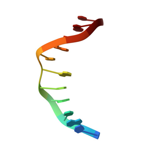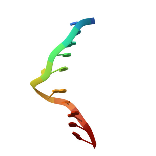
Solution structure of duplex DNA containing the mutagenic lesion 1,N(2)-etheno-2'-deoxyguanine.
Zaliznyak, T., Lukin, M., Johnson, F., de los Santos, C.(2008) Biochemistry 47: 4606-4613
- PubMed: 18373352
- DOI: https://doi.org/10.1021/bi7022514
- Primary Citation of Related Structures:
2K1Y - PubMed Abstract:
We have used high-resolution NMR spectroscopy and molecular dynamics simulations to determine the solution structure of DNA containing the genotoxic lesion 1, N (2)-etheno-2'-deoxyguanosine (epsilonG), paired to dC. The NMR data suggest the presence of a major, minimally perturbed structure at neutral pH. NOESY spectra indicate the presence of a right-handed helix with all nucleotides in anti, 2'-deoxyribose conformations within the C2'-endo/C1'-exo range and proper Watson-Crick base pair alignments outside the lesion site. The epsilonG residue remains deeply embedded inside the helix and stacks between the flanking base pairs. The lesion partner dC is extrahelical and is located in the minor groove of the duplex, where it is highly exposed to solvent. Upon acidification of the sample, a second conformation at the lesion site of the duplex emerges, with protonation of the lesion partner dC and possible formation of a Hoogsteen base pair. Restrained molecular dynamics simulations of the neutral-pH structure generated a set of three-dimensional models that show epsilonG inside the helix, where the lesion is stabilized by stacking interactions with flanking bases but without participating in hydrogen bonding. The lesion counterbase dC is displaced in the minor groove of the duplex where it can form a hydrogen bond with the sugar O4' atom of a residue 2 bp away.
Organizational Affiliation:
Department of Pharmacological Sciences, Stony Brook University, School of Medicine, Stony Brook, NY 11794-8651, USA.