2JE9

CRYSTAL STRUCTURE OF RECOMBINANT DIOCLEA GRANDIFLORA LECTIN COMPLEXED WITH 5-BROMO-4-CHLORO-3-INDOLYL-A-D-MANNOSE


Experimental Data Snapshot

  • Method: X-RAY DIFFRACTION
  • Resolution: 2.10 Å
  • R-Value Free: 0.239 
  • R-Value Work: 0.187 
  • R-Value Observed: 0.190 

wwPDB Validation   3D Report Full Report


Ligand Structure Quality Assessment 


This is version 1.5 of the entry. See complete history


Literature

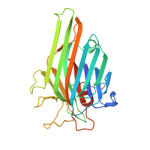
Insights Into the Structural Basis of the Ph- Dependent Dimer-Tetramer Equilibrium Through Crystallographic Analysis of Recombinant Diocleinae Lectins.

Nagano, C.S.Calvete, J.J.Barettino, D.Perez, A.Cavada, B.S.Sanz, L.

(2008) Biochem J 409: 417

  • DOI: https://doi.org/10.1042/BJ20070942
  • Primary Citation of Related Structures:  
    2JDZ, 2JE7, 2JE9, 2JEC

  • PubMed Abstract: 

    The structural ground underlying the pH-dependency of the dimer-tetramer transition of Diocleinae lectins was investigated by equilibrium sedimentation and X-ray crystal structure determination of wild-type and site-directed mutants of recombinant lectins. Synthetic genes coding for the full-length alpha-chains of the seed lectins of Dioclea guianensis (termed r-alphaDguia) and Dioclea grandiflora (termed r-alphaDGL) were designed and expressed in Escherichia coli. This pioneering approach, which will be described in detail in the present paper, yielded recombinant lectins displaying carbohydrate-binding activity, dimer-tetramer equilibria and crystal structures indistinguishable from their natural homologues. Conversion of the pH-stable tetrameric r-alphaDGL into a structure exhibiting pH-dependent dimer-tetramer transition was accomplished through mutations that abolished the interdimeric interactions at the central cavity of the tetrameric lectins. Both the central and the peripheral interacting regions bear structural information for formation of the canonical legume lectin tetramer. We hypothesize that the strength of the ionic contacts at these sites may be modulated by the pH, leading to dissociation of those lectin structures that are not locked into a pH-stable tetramer through interdimeric contacts networking the central cavity loops.


  • Organizational Affiliation

    Instituto de Biomedicina de Valencia, C.S.I.C., Jaime Roig 11, 46010 Valencia, Spain.


Macromolecules
Find similar proteins by:  (by identity cutoff)  |  3D Structure
Entity ID: 1
MoleculeChains Sequence LengthOrganismDetailsImage
LECTIN ALPHA CHAIN
A, B, C, D
239Macropsychanthus grandiflorusMutation(s): 0 
UniProt
Find proteins for P08902 (Dioclea grandiflora)
Explore P08902 
Go to UniProtKB:  P08902
Entity Groups  
Sequence Clusters30% Identity50% Identity70% Identity90% Identity95% Identity100% Identity
UniProt GroupP08902
Sequence Annotations
Expand
  • Reference Sequence
Small Molecules
Ligands 4 Unique
IDChains Name / Formula / InChI Key2D Diagram3D Interactions
XMM
Query on XMM

Download Ideal Coordinates CCD File 
G [auth A],
L [auth B],
R [auth C],
V [auth D]
5-bromo-4-chloro-1H-indol-3-yl alpha-D-mannopyranoside
C14 H15 Br Cl N O6
OPIFSICVWOWJMJ-HAAGFXOZSA-N
SO4
Query on SO4

Download Ideal Coordinates CCD File 
H [auth A]
I [auth A]
M [auth B]
N [auth B]
O [auth B]
H [auth A],
I [auth A],
M [auth B],
N [auth B],
O [auth B],
S [auth C],
W [auth D]
SULFATE ION
O4 S
QAOWNCQODCNURD-UHFFFAOYSA-L
MN
Query on MN

Download Ideal Coordinates CCD File 
F [auth A],
K [auth B],
Q [auth C],
U [auth D]
MANGANESE (II) ION
Mn
WAEMQWOKJMHJLA-UHFFFAOYSA-N
CA
Query on CA

Download Ideal Coordinates CCD File 
E [auth A],
J [auth B],
P [auth C],
T [auth D]
CALCIUM ION
Ca
BHPQYMZQTOCNFJ-UHFFFAOYSA-N
Experimental Data & Validation

Experimental Data

  • Method: X-RAY DIFFRACTION
  • Resolution: 2.10 Å
  • R-Value Free: 0.239 
  • R-Value Work: 0.187 
  • R-Value Observed: 0.190 
  • Space Group: P 21 21 21
Unit Cell:
Length ( Å )Angle ( ˚ )
a = 72.171α = 90
b = 85.153β = 90
c = 175.741γ = 90
Software Package:
Software NamePurpose
REFMACrefinement
MOSFLMdata reduction
SCALEPACKdata scaling
MOLREPphasing

Structure Validation

View Full Validation Report


Ligand Structure Quality Assessment 


Entry History 

Deposition Data

Revision History  (Full details and data files)

  • Version 1.0: 2007-10-30
    Type: Initial release
  • Version 1.1: 2011-05-08
    Changes: Version format compliance
  • Version 1.2: 2011-07-13
    Changes: Version format compliance
  • Version 1.3: 2019-05-08
    Changes: Data collection, Experimental preparation, Other
  • Version 1.4: 2020-07-29
    Type: Remediation
    Reason: Carbohydrate remediation
    Changes: Derived calculations, Other, Structure summary
  • Version 1.5: 2023-12-13
    Changes: Data collection, Database references, Refinement description, Structure summary