2J9D

Structure of GlnK1 with bound effectors indicates regulatory mechanism for ammonia uptake


Experimental Data Snapshot

  • Method: X-RAY DIFFRACTION
  • Resolution: 2.10 Å
  • R-Value Free: 0.265 
  • R-Value Work: 0.207 
  • R-Value Observed: 0.210 

wwPDB Validation   3D Report Full Report


This is version 1.3 of the entry. See complete history


Literature

Structure of Glnk1 with Bound Effectors Indicates Regulatory Mechanism for Ammonia Uptake.

Yildiz, O.Kalthoff, C.Raunser, S.Kuhlbrandt, W.

(2007) EMBO J 26: 589

  • DOI: https://doi.org/10.1038/sj.emboj.7601492
  • Primary Citation of Related Structures:  
    2J9C, 2J9D, 2J9E

  • PubMed Abstract: 

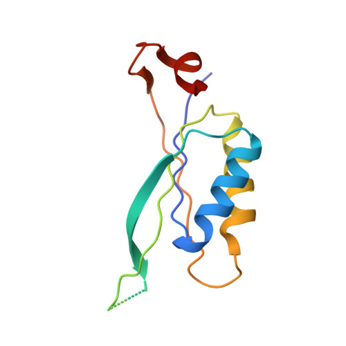
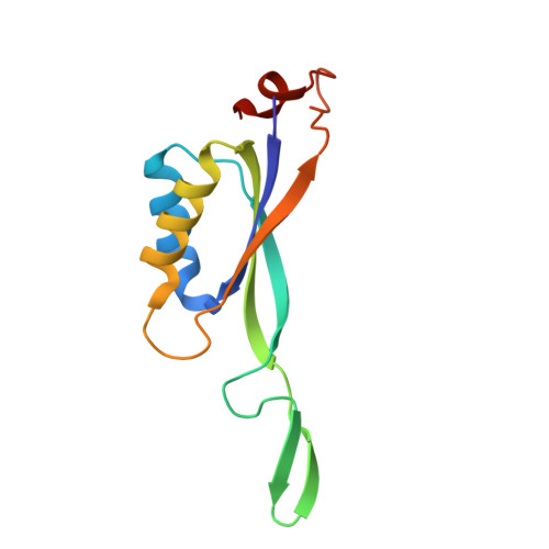
    A binary complex of the ammonia channel Amt1 from Methanococcus jannaschii and its cognate P(II) signalling protein GlnK1 has been produced and characterized. Complex formation is prevented specifically by the effector molecules Mg-ATP and 2-ketoglutarate. Single-particle electron microscopy of the complex shows that GlnK1 binds on the cytoplasmic side of Amt1. Three high-resolution X-ray structures of GlnK1 indicate that the functionally important T-loop has an extended, flexible conformation in the absence of Mg-ATP, but assumes a compact, tightly folded conformation upon Mg-ATP binding, which in turn creates a 2-ketoglutarate-binding site. We propose a regulatory mechanism by which nitrogen uptake is controlled by the binding of both effector molecules to GlnK1. At normal effector levels, a 2-ketoglutarate molecule binding at the apex of the compact T-loop would prevent complex formation, ensuring uninhibited ammonia uptake. At low levels of Mg-ATP, the extended loops would seal the ammonia channels in the complex. Binding of both effector molecules to P(II) signalling proteins may thus represent an effective feedback mechanism for regulating ammonium uptake through the membrane.


  • Organizational Affiliation

    Department of Structural Biology, Max Planck Institute of Biophysics, Frankfurt am Main, Germany.


Macromolecules
Find similar proteins by:  (by identity cutoff)  |  3D Structure
Entity ID: 1
MoleculeChains Sequence LengthOrganismDetailsImage
HYPOTHETICAL NITROGEN REGULATORY PII-LIKE PROTEIN MJ0059
A, B, C, D, F
A, B, C, D, F, G, H, I, J, K, L
119Methanocaldococcus jannaschiiMutation(s): 0 
UniProt
Find proteins for Q60381 (Methanocaldococcus jannaschii (strain ATCC 43067 / DSM 2661 / JAL-1 / JCM 10045 / NBRC 100440))
Explore Q60381 
Go to UniProtKB:  Q60381
Entity Groups  
Sequence Clusters30% Identity50% Identity70% Identity90% Identity95% Identity100% Identity
UniProt GroupQ60381
Sequence Annotations
Expand
  • Reference Sequence
Find similar proteins by:  (by identity cutoff)  |  3D Structure
Entity ID: 2
MoleculeChains Sequence LengthOrganismDetailsImage
HYPOTHETICAL NITROGEN REGULATORY PII-LIKE PROTEIN MJ0059119Methanocaldococcus jannaschiiMutation(s): 0 
UniProt
Find proteins for Q60381 (Methanocaldococcus jannaschii (strain ATCC 43067 / DSM 2661 / JAL-1 / JCM 10045 / NBRC 100440))
Explore Q60381 
Go to UniProtKB:  Q60381
Entity Groups  
Sequence Clusters30% Identity50% Identity70% Identity90% Identity95% Identity100% Identity
UniProt GroupQ60381
Sequence Annotations
Expand
  • Reference Sequence
Small Molecules
Ligands 4 Unique
IDChains Name / Formula / InChI Key2D Diagram3D Interactions
ADP
Query on ADP

Download Ideal Coordinates CCD File 
O [auth B],
S [auth I],
U [auth J],
W [auth L]
ADENOSINE-5'-DIPHOSPHATE
C10 H15 N5 O10 P2
XTWYTFMLZFPYCI-KQYNXXCUSA-N
AMP
Query on AMP

Download Ideal Coordinates CCD File 
P [auth E]ADENOSINE MONOPHOSPHATE
C10 H14 N5 O7 P
UDMBCSSLTHHNCD-KQYNXXCUSA-N
ACT
Query on ACT

Download Ideal Coordinates CCD File 
M [auth A],
N [auth A],
Q [auth E],
R [auth H],
V [auth J]
ACETATE ION
C2 H3 O2
QTBSBXVTEAMEQO-UHFFFAOYSA-M
CL
Query on CL

Download Ideal Coordinates CCD File 
T [auth J]CHLORIDE ION
Cl
VEXZGXHMUGYJMC-UHFFFAOYSA-M
Experimental Data & Validation

Experimental Data

  • Method: X-RAY DIFFRACTION
  • Resolution: 2.10 Å
  • R-Value Free: 0.265 
  • R-Value Work: 0.207 
  • R-Value Observed: 0.210 
  • Space Group: P 21 21 21
Unit Cell:
Length ( Å )Angle ( ˚ )
a = 96.6α = 90
b = 107.03β = 90
c = 134.34γ = 90
Software Package:
Software NamePurpose
REFMACrefinement
XDSdata reduction
XSCALEdata scaling
PHASERphasing

Structure Validation

View Full Validation Report


Entry History 

Deposition Data

Revision History  (Full details and data files)

  • Version 1.0: 2007-01-16
    Type: Initial release
  • Version 1.1: 2011-05-08
    Changes: Version format compliance
  • Version 1.2: 2011-07-13
    Changes: Version format compliance
  • Version 1.3: 2023-12-13
    Changes: Data collection, Database references, Refinement description