2J8U

Large CDR3a loop alteration as a function of MHC mutation.


Experimental Data Snapshot

  • Method: X-RAY DIFFRACTION
  • Resolution: 2.88 Å
  • R-Value Free: 0.293 
  • R-Value Work: 0.268 
  • R-Value Observed: 0.269 

wwPDB Validation   3D Report Full Report


This is version 1.4 of the entry. See complete history


Literature

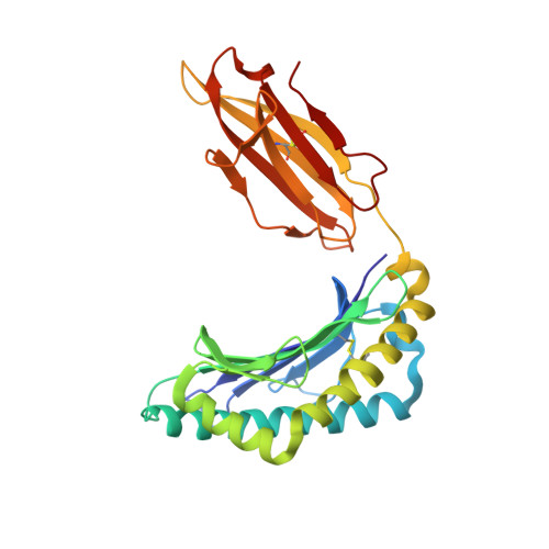
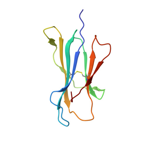
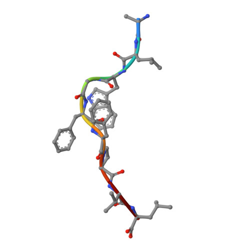
Single MHC mutation eliminates enthalpy associated with T cell receptor binding.

Miller, P.J.Pazy, Y.Conti, B.Riddle, D.Appella, E.Collins, E.J.

(2007) J Mol Biol 373: 315-327

  • DOI: https://doi.org/10.1016/j.jmb.2007.07.028
  • Primary Citation of Related Structures:  
    2J8U, 2JCC, 2UWE

  • PubMed Abstract: 

    The keystone of the adaptive immune response is T cell receptor (TCR) recognition of peptide presented by major histocompatibility complex (pMHC) molecules. The crystal structure of AHIII TCR bound to MHC, HLA-A2, showed a large interface with an atypical binding orientation. MHC mutations in the interface of the proteins were tested for changes in TCR recognition. From the range of responses observed, three representative HLA-A2 mutants, T163A, W167A, and K66A, were selected for further study. Binding constants and co-crystal structures of the AHIII TCR and the three mutants were determined. K66 in HLA-A2 makes contacts with both peptide and TCR, and has been identified as a critical residue for recognition by numerous TCR. The K66A mutation resulted in the lowest AHIII T cell response and the lowest binding affinity, which suggests that the T cell response may correlate with affinity. Importantly, the K66A mutation does not affect the conformation of the peptide. The change in affinity appears to be due to a loss in hydrogen bonds in the interface as a result of a conformational change in the TCR complementarity-determining region 3 (CDR3) loop. Isothermal titration calorimetry confirmed the loss of hydrogen bonding by a large loss in enthalpy. Our findings are inconsistent with the notion that the CDR1 and CDR2 loops of the TCR are responsible for MHC restriction, while the CDR3 loops interact solely with the peptide. Instead, we present here an MHC mutation that does not change the conformation of the peptide, yet results in an altered conformation of a CDR3.


  • Organizational Affiliation

    Department of Biochemistry and Biophysics, The University of North Carolina at Chapel Hill, Chapel Hill, NC 27599, USA.


Macromolecules
Find similar proteins by:  (by identity cutoff)  |  3D Structure
Entity ID: 1
MoleculeChains Sequence LengthOrganismDetailsImage
HLA class I histocompatibility antigen, A-2 alpha chainA,
F [auth H]
275Homo sapiensMutation(s): 1 
Gene Names: HLA-AHLAA
UniProt & NIH Common Fund Data Resources
Find proteins for P04439 (Homo sapiens)
Explore P04439 
Go to UniProtKB:  P04439
PHAROS:  P04439
GTEx:  ENSG00000206503 
Entity Groups  
Sequence Clusters30% Identity50% Identity70% Identity90% Identity95% Identity100% Identity
UniProt GroupP04439
Sequence Annotations
Expand
  • Reference Sequence
Find similar proteins by:  (by identity cutoff)  |  3D Structure
Entity ID: 2
MoleculeChains Sequence LengthOrganismDetailsImage
Beta-2-microglobulinB,
G [auth I]
100Homo sapiensMutation(s): 0 
Gene Names: B2MCDABP0092HDCMA22P
UniProt & NIH Common Fund Data Resources
Find proteins for P61769 (Homo sapiens)
Explore P61769 
Go to UniProtKB:  P61769
PHAROS:  P61769
GTEx:  ENSG00000166710 
Entity Groups  
Sequence Clusters30% Identity50% Identity70% Identity90% Identity95% Identity100% Identity
UniProt GroupP61769
Sequence Annotations
Expand
  • Reference Sequence

Find similar proteins by:  Sequence   |   3D Structure  

Entity ID: 3
MoleculeChains Sequence LengthOrganismDetailsImage
SELF-PEPTIDE P1049C,
H [auth J]
9Homo sapiensMutation(s): 0 
UniProt & NIH Common Fund Data Resources
Find proteins for Q9NPA0 (Homo sapiens)
Explore Q9NPA0 
Go to UniProtKB:  Q9NPA0
PHAROS:  Q9NPA0
GTEx:  ENSG00000134153 
Entity Groups  
UniProt GroupQ9NPA0
Sequence Annotations
Expand
  • Reference Sequence
Find similar proteins by:  (by identity cutoff)  |  3D Structure
Entity ID: 4
MoleculeChains Sequence LengthOrganismDetailsImage
AHIII TCR ALPHA CHAIND [auth E],
I [auth L]
194Mus musculusMutation(s): 0 
UniProt
Find proteins for P01849 (Mus musculus)
Explore P01849 
Go to UniProtKB:  P01849
Entity Groups  
Sequence Clusters30% Identity50% Identity70% Identity90% Identity95% Identity100% Identity
UniProt GroupP01849
Sequence Annotations
Expand
  • Reference Sequence
Find similar proteins by:  (by identity cutoff)  |  3D Structure
Entity ID: 5
MoleculeChains Sequence LengthOrganismDetailsImage
AHIII TCR BETA CHAINE [auth F],
J [auth M]
238Mus musculusMutation(s): 0 
UniProt
Find proteins for P04213 (Mus musculus)
Explore P04213 
Go to UniProtKB:  P04213
Entity Groups  
Sequence Clusters30% Identity50% Identity70% Identity90% Identity95% Identity100% Identity
UniProt GroupP04213
Sequence Annotations
Expand
  • Reference Sequence
Experimental Data & Validation

Experimental Data

  • Method: X-RAY DIFFRACTION
  • Resolution: 2.88 Å
  • R-Value Free: 0.293 
  • R-Value Work: 0.268 
  • R-Value Observed: 0.269 
  • Space Group: P 1 21 1
Unit Cell:
Length ( Å )Angle ( ˚ )
a = 93.422α = 90
b = 83.893β = 92.21
c = 122.273γ = 90
Software Package:
Software NamePurpose
REFMACrefinement
HKL-2000data reduction
HKL-2000data scaling
PHASERphasing

Structure Validation

View Full Validation Report


Entry History 

Deposition Data

Revision History  (Full details and data files)

  • Version 1.0: 2007-10-16
    Type: Initial release
  • Version 1.1: 2011-07-13
    Changes: Advisory, Version format compliance
  • Version 1.2: 2011-08-03
    Changes: Derived calculations, Other, Refinement description, Source and taxonomy, Structure summary
  • Version 1.3: 2018-12-12
    Changes: Advisory, Data collection, Database references, Source and taxonomy, Structure summary
  • Version 1.4: 2023-12-13
    Changes: Advisory, Data collection, Database references, Other, Refinement description