2J6E

Crystal Structure of an Autoimmune Complex between a Human IgM Rheumatoid Factor and IgG1 Fc reveals a Novel Fc Epitope and Evidence for Affinity Maturation


Experimental Data Snapshot

  • Method: X-RAY DIFFRACTION
  • Resolution: 3.00 Å
  • R-Value Free: 0.288 
  • R-Value Work: 0.223 
  • R-Value Observed: 0.223 

wwPDB Validation   3D Report Full Report


This is version 4.1 of the entry. See complete history


Literature

Crystal structure of a human autoimmune complex between IgM rheumatoid factor RF61 and IgG1 Fc reveals a novel epitope and evidence for affinity maturation.

Duquerroy, S.Stura, E.A.Bressanelli, S.Fabiane, S.M.Vaney, M.C.Beale, D.Hamon, M.Casali, P.Rey, F.A.Sutton, B.J.Taussig, M.J.

(2007) J Mol Biol 368: 1321-1331

  • DOI: https://doi.org/10.1016/j.jmb.2007.02.085
  • Primary Citation of Related Structures:  
    2J6E

  • PubMed Abstract: 

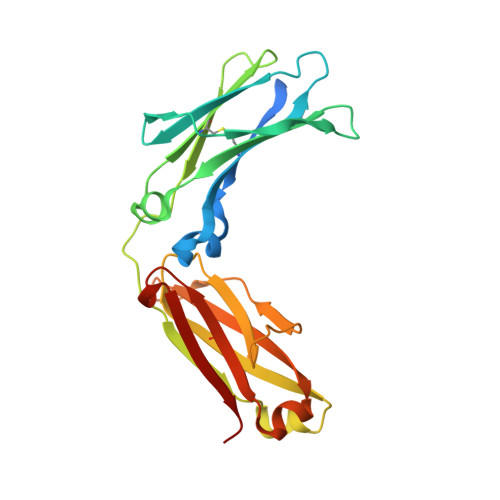
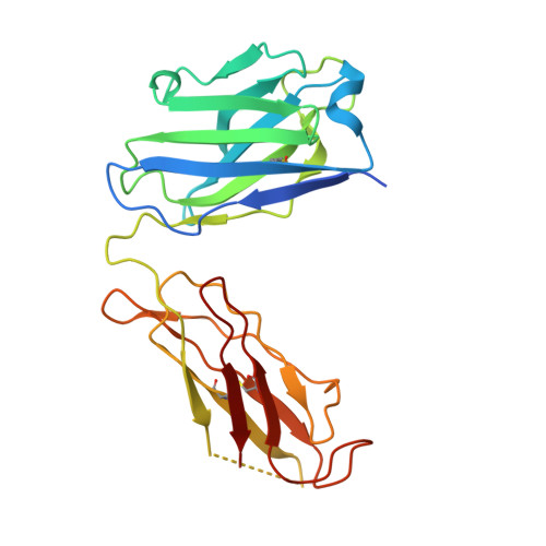
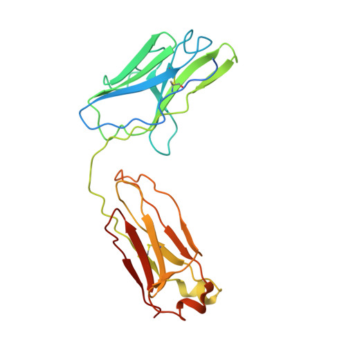
    Rheumatoid factors (RF) are autoantibodies that recognize epitopes in the Fc region of immunoglobulin (Ig) G and that correlate with the clinical severity of rheumatoid arthritis (RA). Here we report the X-ray crystallographic structure, at 3 A resolution, of a complex between the Fc region of human IgG1 and the Fab fragment of a monoclonal IgM RF (RF61), derived from an RA patient and with a relatively high affinity for IgG Fc. In the complex, two Fab fragments bind to each Fc at epitopes close to the C terminus, and each epitope comprises residues from both Cgamma3 domains. A central role in the unusually hydrophilic epitope is played by the side-chain of Arg355, accounting for the subclass specificity of RF61, which recognizes IgG1,-2, and -3 in preference to IgG4, in which the corresponding residue is Gln355. Compared with a previously determined complex of a lower affinity RF (RF-AN) bound to IgG4 Fc, in which only residues at the very edge of the antibody combining site were involved in binding, the epitope bound by RF61 is centered in classic fashion on the axis of the V(H):V(L) beta-barrel. The complementarity determining region-H3 loop plays a key role, forming a pocket in which Arg355 is bound by two salt-bridges. The antibody contacts also involve two somatically mutated V(H) residues, reinforcing the suggestion of a process of antigen-driven maturation and selection for IgG Fc during the generation of this RF autoantibody.


  • Organizational Affiliation

    Virologie Moléculaire et Structurale, CNRS UMR 2472-INRA UMR 1157, 91198 Gif-sur-Yvette, France.


Macromolecules
Find similar proteins by:  (by identity cutoff)  |  3D Structure
Entity ID: 1
MoleculeChains Sequence LengthOrganismDetailsImage
IG GAMMA-1 CHAIN C REGION
A, B
232Homo sapiensMutation(s): 0 
UniProt & NIH Common Fund Data Resources
Find proteins for P01857 (Homo sapiens)
Explore P01857 
Go to UniProtKB:  P01857
PHAROS:  P01857
GTEx:  ENSG00000211896 
Entity Groups  
Sequence Clusters30% Identity50% Identity70% Identity90% Identity95% Identity100% Identity
UniProt GroupP01857
Sequence Annotations
Expand
  • Reference Sequence
Find similar proteins by:  (by identity cutoff)  |  3D Structure
Entity ID: 2
MoleculeChains Sequence LengthOrganismDetailsImage
IGMC [auth H],
D [auth I]
231Homo sapiensMutation(s): 0 
Entity Groups  
Sequence Clusters30% Identity50% Identity70% Identity90% Identity95% Identity100% Identity
Sequence Annotations
Expand
  • Reference Sequence
Find similar proteins by:  (by identity cutoff)  |  3D Structure
Entity ID: 3
MoleculeChains Sequence LengthOrganismDetailsImage
IGME [auth L],
F [auth M]
234Homo sapiensMutation(s): 0 
Entity Groups  
Sequence Clusters30% Identity50% Identity70% Identity90% Identity95% Identity100% Identity
Sequence Annotations
Expand
  • Reference Sequence
Oligosaccharides

Help

Entity ID: 4
MoleculeChains Length2D Diagram Glycosylation3D Interactions
beta-D-galactopyranose-(1-4)-2-acetamido-2-deoxy-beta-D-glucopyranose-(1-2)-alpha-D-mannopyranose-(1-6)-[2-acetamido-2-deoxy-beta-D-glucopyranose-(1-2)-beta-D-mannopyranose-(1-3)]beta-D-mannopyranose-(1-4)-2-acetamido-2-deoxy-beta-D-glucopyranose-(1-4)-[beta-L-fucopyranose-(1-6)]2-acetamido-2-deoxy-beta-D-glucopyranoseG [auth C],
H [auth D]
9N-Glycosylation
Glycosylation Resources
GlyTouCan:  G19282BU
GlyCosmos:  G19282BU
GlyGen:  G19282BU
Small Molecules
Ligands 5 Unique
IDChains Name / Formula / InChI Key2D Diagram3D Interactions
CAC
Query on CAC

Download Ideal Coordinates CCD File 
K [auth A]CACODYLATE ION
C2 H6 As O2
OGGXGZAMXPVRFZ-UHFFFAOYSA-M
MPD
Query on MPD

Download Ideal Coordinates CCD File 
L [auth A]
M [auth A]
N [auth A]
R [auth B]
S [auth B]
L [auth A],
M [auth A],
N [auth A],
R [auth B],
S [auth B],
V [auth M]
(4S)-2-METHYL-2,4-PENTANEDIOL
C6 H14 O2
SVTBMSDMJJWYQN-YFKPBYRVSA-N
CD
Query on CD

Download Ideal Coordinates CCD File 
I [auth A]CADMIUM ION
Cd
WLZRMCYVCSSEQC-UHFFFAOYSA-N
ZN
Query on ZN

Download Ideal Coordinates CCD File 
J [auth A],
Q [auth B]
ZINC ION
Zn
PTFCDOFLOPIGGS-UHFFFAOYSA-N
ACT
Query on ACT

Download Ideal Coordinates CCD File 
O [auth A],
P [auth A],
T [auth B],
U [auth H]
ACETATE ION
C2 H3 O2
QTBSBXVTEAMEQO-UHFFFAOYSA-M
Experimental Data & Validation

Experimental Data

  • Method: X-RAY DIFFRACTION
  • Resolution: 3.00 Å
  • R-Value Free: 0.288 
  • R-Value Work: 0.223 
  • R-Value Observed: 0.223 
  • Space Group: C 1 2 1
Unit Cell:
Length ( Å )Angle ( ˚ )
a = 241.98α = 90
b = 75.61β = 91.13
c = 102.4γ = 90
Software Package:
Software NamePurpose
CNSrefinement
DENZOdata reduction
SCALEPACKdata scaling
AMoREphasing

Structure Validation

View Full Validation Report


Entry History 

Deposition Data

Revision History  (Full details and data files)

  • Version 1.0: 2007-04-10
    Type: Initial release
  • Version 1.1: 2011-05-08
    Changes: Version format compliance
  • Version 1.2: 2011-07-13
    Changes: Version format compliance
  • Version 1.3: 2019-04-03
    Changes: Data collection, Derived calculations, Other, Source and taxonomy
  • Version 1.4: 2019-08-14
    Changes: Data collection, Database references, Experimental preparation, Structure summary
  • Version 2.0: 2020-07-29
    Type: Remediation
    Reason: Carbohydrate remediation
    Changes: Atomic model, Data collection, Derived calculations, Other, Structure summary
  • Version 3.0: 2022-05-04
    Changes: Atomic model, Data collection, Database references, Derived calculations, Structure summary
  • Version 4.0: 2022-05-11
    Changes: Advisory, Atomic model, Data collection, Derived calculations, Non-polymer description, Structure summary
  • Version 4.1: 2023-12-13
    Changes: Data collection, Refinement description