2J4Y

Crystal structure of a rhodopsin stabilizing mutant expressed in mammalian cells


Experimental Data Snapshot

  • Method: X-RAY DIFFRACTION
  • Resolution: 3.40 Å
  • R-Value Free: 0.330 
  • R-Value Work: 0.290 
  • R-Value Observed: 0.290 

wwPDB Validation   3D Report Full Report


This is version 1.5 of the entry. See complete history

Re-refinement Note

A newer entry is available that reflects an alternative modeling of the original data: 3C9M


Literature

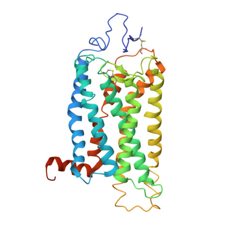
Crystal Structure of a Thermally Stable Rhodopsin Mutant.

Standfuss, J.Xie, G.Edwards, P.C.Burghammer, M.Oprian, D.D.Schertler, G.F.X.

(2007) J Mol Biol 372: 1179

  • DOI: https://doi.org/10.1016/j.jmb.2007.03.007
  • Primary Citation of Related Structures:  
    2J4Y

  • PubMed Abstract: 

    We determined the structure of the rhodopsin mutant N2C/D282C expressed in mammalian cells; the first structure of a recombinantly produced G protein-coupled receptor (GPCR). The mutant was designed to form a disulfide bond between the N terminus and loop E3, which allows handling of opsin in detergent solution and increases thermal stability of rhodopsin by 10 deg.C. It allowed us to crystallize a fully deglycosylated rhodopsin (N2C/N15D/D282C). N15 mutations are normally misfolding and cause retinitis pigmentosa in humans. Microcrystallographic techniques and a 5 microm X-ray beam were used to collect data along a single needle measuring 5 microm x 5 microm x 90 microm. The disulfide introduces only minor changes but fixes the N-terminal cap over the beta-sheet lid covering the ligand-binding site, a likely explanation for the increased stability. This work allows structural investigation of rhodopsin mutants and shows the problems encountered during structure determination of GPCRs and other mammalian membrane proteins.


  • Organizational Affiliation

    MRC Laboratory of Molecular Biology, Structural Studies, Hills Road, Cambridge CB2 2QH, UK.


Macromolecules
Find similar proteins by:  (by identity cutoff)  |  3D Structure
Entity ID: 1
MoleculeChains Sequence LengthOrganismDetailsImage
RHODOPSIN
A, B
349Bos taurusMutation(s): 3 
Membrane Entity: Yes 
UniProt
Find proteins for P02699 (Bos taurus)
Explore P02699 
Go to UniProtKB:  P02699
Entity Groups  
Sequence Clusters30% Identity50% Identity70% Identity90% Identity95% Identity100% Identity
UniProt GroupP02699
Sequence Annotations
Expand
  • Reference Sequence
Experimental Data & Validation

Experimental Data

  • Method: X-RAY DIFFRACTION
  • Resolution: 3.40 Å
  • R-Value Free: 0.330 
  • R-Value Work: 0.290 
  • R-Value Observed: 0.290 
  • Space Group: P 31
Unit Cell:
Length ( Å )Angle ( ˚ )
a = 109.3α = 90
b = 109.3β = 90
c = 77.7γ = 120
Software Package:
Software NamePurpose
CNSrefinement
XDSdata reduction
XSCALEdata scaling
MOLREPphasing

Structure Validation

View Full Validation Report


Entry History 

Deposition Data

Revision History  (Full details and data files)

  • Version 1.0: 2007-09-25
    Type: Initial release
  • Version 1.1: 2012-12-12
    Changes: Atomic model, Database references, Derived calculations, Non-polymer description, Other, Refinement description, Source and taxonomy, Structure summary, Version format compliance
  • Version 1.2: 2019-05-08
    Changes: Data collection, Derived calculations, Experimental preparation, Other
  • Version 1.3: 2019-10-23
    Changes: Data collection, Database references, Other
  • Version 1.4: 2020-07-29
    Type: Remediation
    Reason: Carbohydrate remediation
    Changes: Data collection, Derived calculations, Structure summary
  • Version 1.5: 2023-12-13
    Changes: Data collection, Database references, Refinement description, Structure summary