2J08

Thermus DNA photolyase with 8-Iod-riboflavin antenna chromophore


Experimental Data Snapshot

  • Method: X-RAY DIFFRACTION
  • Resolution: 2.61 Å
  • R-Value Free: 0.256 
  • R-Value Work: 0.163 
  • R-Value Observed: 0.167 

wwPDB Validation   3D Report Full Report


Ligand Structure Quality Assessment 


This is version 1.3 of the entry. See complete history


Literature

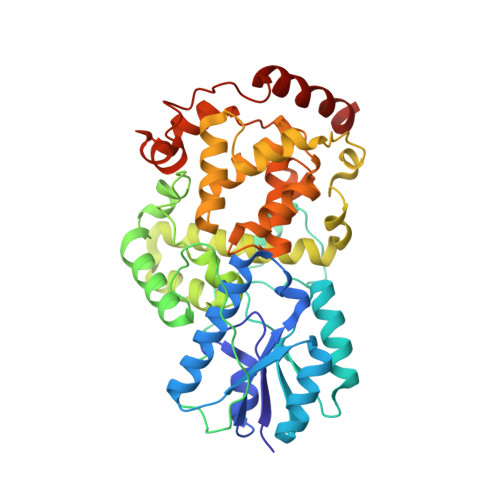
Natural and Non-Natural Antenna Chromophores in the DNA Photolyase from Thermus Thermophilus

Klar, T.Kaiser, G.Hennecke, U.Carell, T.Batschauer, A.Essen, L.-O.

(2006) Chembiochem 7: 1798

  • DOI: https://doi.org/10.1002/cbic.200600206
  • Primary Citation of Related Structures:  
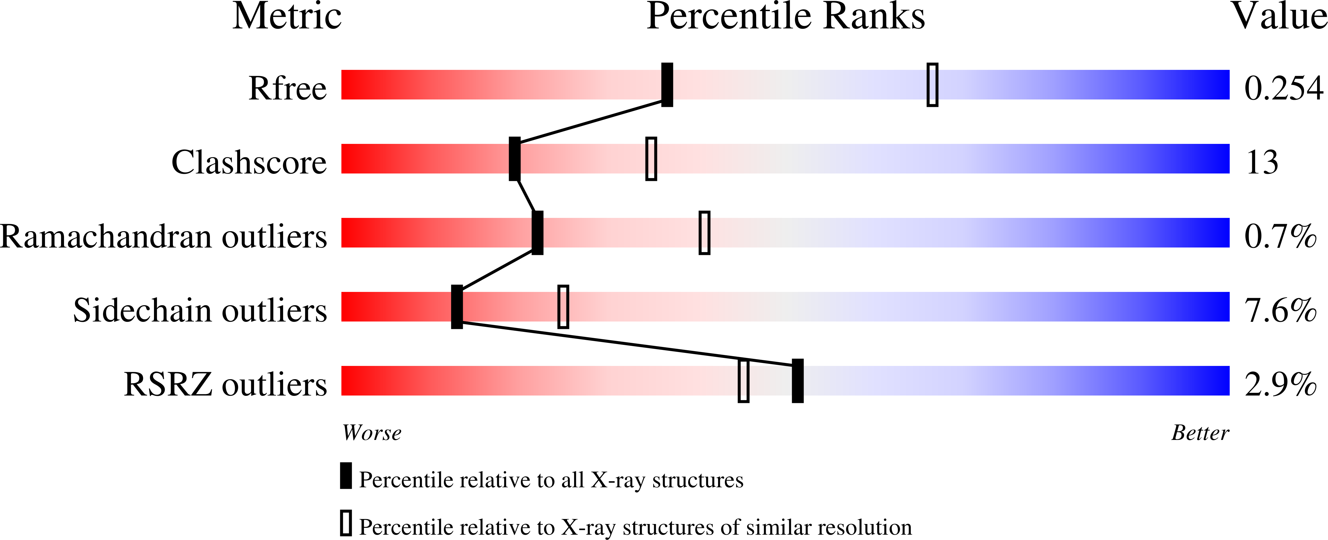
    2J07, 2J08, 2J09

  • PubMed Abstract: 

    X-ray crystallographic and functional analysis of the class I DNA photolyase from Thermus thermophilus revealed the binding of flavin mononucleotide (FMN) as an antenna chromophore. The binding mode of FMN closely coincides with the binding of a deazaflavin-like chromophore in the related class I DNA photolyase from Anacystis nidulans. Compared to the R46E mutant, which lacks a conserved arginine in the binding site for the antenna chromophore, the FMN-comprising holophotolyase exhibits an eightfold higher activity at 450 nm. The facile incorporation of the flavin cofactors 8-hydroxy-deazariboflavin and 8-iodo-8-demethyl-riboflavin into the binding site for the antenna chromophore paves the way for wavelength-tuning of the activity spectra of DNA photolyases by using synthetic flavins.


  • Organizational Affiliation

    Philipps-Universität Marburg, Fachbereich Chemie, Hans-Meerwein-Strasse, 35032 Marburg, Germany.


Macromolecules
Find similar proteins by:  (by identity cutoff)  |  3D Structure
Entity ID: 1
MoleculeChains Sequence LengthOrganismDetailsImage
DEOXYRIBODIPYRIMIDINE PHOTO-LYASE420Thermus thermophilus HB8Mutation(s): 0 
EC: 4.1.99.3
UniProt
Find proteins for P61497 (Thermus thermophilus (strain ATCC 27634 / DSM 579 / HB8))
Explore P61497 
Go to UniProtKB:  P61497
Entity Groups  
Sequence Clusters30% Identity50% Identity70% Identity90% Identity95% Identity100% Identity
UniProt GroupP61497
Sequence Annotations
Expand
  • Reference Sequence
Small Molecules
Ligands 4 Unique
IDChains Name / Formula / InChI Key2D Diagram3D Interactions
FAD
Query on FAD

Download Ideal Coordinates CCD File 
D [auth A]FLAVIN-ADENINE DINUCLEOTIDE
C27 H33 N9 O15 P2
VWWQXMAJTJZDQX-UYBVJOGSSA-N
IRF
Query on IRF

Download Ideal Coordinates CCD File 
E [auth A]1-DEOXY-1-(8-IODO-7-METHYL-2,4-DIOXO-3,4-DIHYDROBENZO[G]PTERIDIN-10(2H)-YL)-D-RIBITOL
C16 H17 I N4 O6
GXUZMSUHTJJUNU-LOWVWBTDSA-N
PO4
Query on PO4

Download Ideal Coordinates CCD File 
F [auth A],
G [auth A]
PHOSPHATE ION
O4 P
NBIIXXVUZAFLBC-UHFFFAOYSA-K
CL
Query on CL

Download Ideal Coordinates CCD File 
B [auth A],
C [auth A]
CHLORIDE ION
Cl
VEXZGXHMUGYJMC-UHFFFAOYSA-M
Experimental Data & Validation

Experimental Data

  • Method: X-RAY DIFFRACTION
  • Resolution: 2.61 Å
  • R-Value Free: 0.256 
  • R-Value Work: 0.163 
  • R-Value Observed: 0.167 
  • Space Group: P 61 2 2
Unit Cell:
Length ( Å )Angle ( ˚ )
a = 112.62α = 90
b = 112.62β = 90
c = 140.262γ = 120
Software Package:
Software NamePurpose
REFMACrefinement
MOSFLMdata reduction
SCALAdata scaling
MOLREPphasing

Structure Validation

View Full Validation Report


Ligand Structure Quality Assessment 


Entry History 

Deposition Data

Revision History  (Full details and data files)

  • Version 1.0: 2007-05-01
    Type: Initial release
  • Version 1.1: 2011-07-13
    Changes: Advisory, Version format compliance
  • Version 1.2: 2017-07-05
    Changes: Data collection
  • Version 1.3: 2023-12-13
    Changes: Data collection, Database references, Derived calculations, Other, Refinement description