2J05

Crystal structure of the RasGAP SH3 domain at 1.5 Angstrom resolution


Experimental Data Snapshot

  • Method: X-RAY DIFFRACTION
  • Resolution: 1.50 Å
  • R-Value Free: 0.199 
  • R-Value Work: 0.177 
  • R-Value Observed: 0.177 

wwPDB Validation   3D Report Full Report


This is version 1.2 of the entry. See complete history


Literature

High Resolution Crystal Structures of the P120 Rasgap SH3 Domain.

Ross, B.Kristensen, O.Favre, D.Walicki, J.Kastrup, J.S.Widmann, C.Gajhede, M.

(2007) Biochem Biophys Res Commun 353: 463

  • DOI: https://doi.org/10.1016/j.bbrc.2006.12.044
  • Primary Citation of Related Structures:  
    2J05, 2J06

  • PubMed Abstract: 

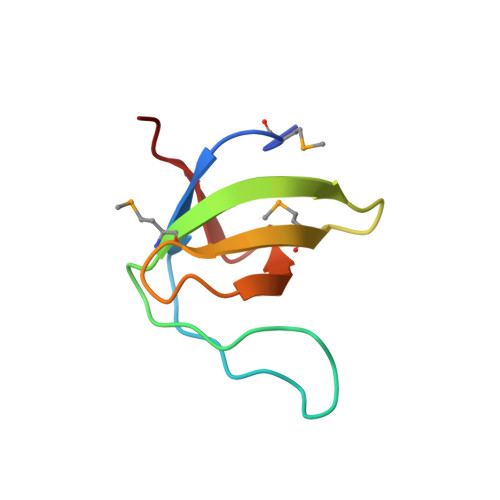
    X-ray structures of two crystal forms of the Src homology 3 domain (SH3) of the Ras GTPase activating protein (RasGAP) were determined at 1.5 and 1.8A resolution. The overall structure comprises a single domain with two tightly packed beta-sheets linked by a short helical segment. An important motif for peptide binding in other SH3 domains is not conserved in RasGAP. The RasGAP SH3 domain forms dimers in the crystal structures, which may provide new functional insight. The dimer interface involves residues also present in a peptide previously identified as an apoptotic sensitizer of tumor cells.


  • Organizational Affiliation

    Biostructural Research, Department of Medicinal Chemistry, The Danish University of Pharmaceutical Sciences, Universitetsparken 2, DK-2100 Copenhagen, Denmark.


Macromolecules
Find similar proteins by:  (by identity cutoff)  |  3D Structure
Entity ID: 1
MoleculeChains Sequence LengthOrganismDetailsImage
RAS GTPASE-ACTIVATING PROTEIN 1
A, B
65Homo sapiensMutation(s): 0 
UniProt & NIH Common Fund Data Resources
Find proteins for P20936 (Homo sapiens)
Explore P20936 
Go to UniProtKB:  P20936
PHAROS:  P20936
GTEx:  ENSG00000145715 
Entity Groups  
Sequence Clusters30% Identity50% Identity70% Identity90% Identity95% Identity100% Identity
UniProt GroupP20936
Sequence Annotations
Expand
  • Reference Sequence
Small Molecules
Modified Residues  1 Unique
IDChains TypeFormula2D DiagramParent
MSE
Query on MSE
A, B
L-PEPTIDE LINKINGC5 H11 N O2 SeMET
Experimental Data & Validation

Experimental Data

  • Method: X-RAY DIFFRACTION
  • Resolution: 1.50 Å
  • R-Value Free: 0.199 
  • R-Value Work: 0.177 
  • R-Value Observed: 0.177 
  • Space Group: P 61
Unit Cell:
Length ( Å )Angle ( ˚ )
a = 32.8α = 90
b = 32.8β = 90
c = 183.33γ = 120
Software Package:
Software NamePurpose
CNSrefinement
XDSdata reduction
XDSdata scaling

Structure Validation

View Full Validation Report


Entry History 

Deposition Data

Revision History  (Full details and data files)

  • Version 1.0: 2007-01-02
    Type: Initial release
  • Version 1.1: 2011-05-08
    Changes: Version format compliance
  • Version 1.2: 2011-07-13
    Changes: Version format compliance