2IWF

Resting form of pink nitrous oxide reductase from Achromobacter Cycloclastes


Experimental Data Snapshot

  • Method: X-RAY DIFFRACTION
  • Resolution: 1.86 Å
  • R-Value Free: 0.271 
  • R-Value Work: 0.209 
  • R-Value Observed: 0.209 

wwPDB Validation   3D Report Full Report


This is version 1.2 of the entry. See complete history


Literature

Insight Into Catalysis of Nitrous Oxide Reductase from High-Resolution Structures of Resting and Inhibitor-Bound Enzyme from Achromobacter Cycloclastes.

Paraskevopoulos, K.Antonyuk, S.V.Sawers, R.G.Eady, R.R.Hasnain, S.S.

(2006) J Mol Biol 362: 55

  • DOI: https://doi.org/10.1016/j.jmb.2006.06.064
  • Primary Citation of Related Structures:  
    2IWF, 2IWK

  • PubMed Abstract: 

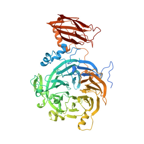
    The difficult chemistry of nitrous oxide (N2O) reduction to gaseous nitrogen (N2) in biology is catalysed by the novel micro4-sulphide-bridged tetranuclear Cuz cluster of the N2O reductases (N2OR). Two spectroscopically distinct forms of this cluster have been identified as CuZ and CuZ*. We have obtained a 1.86 A resolution crystal structure of the pink-purple species of N2OR from Achromobacter cycloclastes (AcN2OR) isolated under aerobic conditions. This structure reveals a previously unobserved ligation with two oxygen atoms from H2O/OH- coordinated to Cu1 and Cu4 of the catalytic centre. We ascribe this structure to be that of the CuZ form of the cluster, since the previously reported structures of two blue species of N2ORs, also isolated aerobically, have characterised the redox inactive CuZ* form, revealing a single water molecule at Cu4. Exposure of the as-isolated AcN2OR to sodium iodide led to reduction of the electron-donating CuA site and the formation of a blue species. Structure determination of this adduct at 1.7 A resolution showed that iodide was bound at the CuZ site bridging the Cu1 and Cu4 ions. This structure represents the first observation of an inhibitor bound to the Cu1-Cu4 edge of the catalytic cluster, providing clear evidence for this being the catalytic edge in N2ORs. These structures, together with the published structural and spectroscopic data, give fresh insight into the mode of substrate binding, reduction and catalysis.


  • Organizational Affiliation

    School of Biomolecular Sciences, Liverpool John Moores University, Liverpool L3 5AF, UK.


Macromolecules
Find similar proteins by:  (by identity cutoff)  |  3D Structure
Entity ID: 1
MoleculeChains Sequence LengthOrganismDetailsImage
NITROUS-OXIDE REDUCTASE
A, B
642Achromobacter cycloclastesMutation(s): 0 
EC: 1.7.99.6
UniProt
Find proteins for P94127 (Achromobacter cycloclastes)
Explore P94127 
Go to UniProtKB:  P94127
Entity Groups  
Sequence Clusters30% Identity50% Identity70% Identity90% Identity95% Identity100% Identity
UniProt GroupP94127
Sequence Annotations
Expand
  • Reference Sequence
Small Molecules
Ligands 5 Unique
IDChains Name / Formula / InChI Key2D Diagram3D Interactions
CUZ
Query on CUZ

Download Ideal Coordinates CCD File 
E [auth A],
R [auth B]
(MU-4-SULFIDO)-TETRA-NUCLEAR COPPER ION
Cu4 S
IGIWMDIIFLPFOP-UHFFFAOYSA-N
CU
Query on CU

Download Ideal Coordinates CCD File 
C [auth A],
D [auth A],
P [auth B],
Q [auth B]
COPPER (II) ION
Cu
JPVYNHNXODAKFH-UHFFFAOYSA-N
CA
Query on CA

Download Ideal Coordinates CCD File 
F [auth A],
G [auth A],
H [auth A],
S [auth B],
T [auth B]
CALCIUM ION
Ca
BHPQYMZQTOCNFJ-UHFFFAOYSA-N
CL
Query on CL

Download Ideal Coordinates CCD File 
I [auth A],
U [auth B]
CHLORIDE ION
Cl
VEXZGXHMUGYJMC-UHFFFAOYSA-M
NA
Query on NA

Download Ideal Coordinates CCD File 
AA [auth B]
BA [auth B]
J [auth A]
K [auth A]
L [auth A]
AA [auth B],
BA [auth B],
J [auth A],
K [auth A],
L [auth A],
M [auth A],
N [auth A],
O [auth A],
V [auth B],
W [auth B],
X [auth B],
Y [auth B],
Z [auth B]
SODIUM ION
Na
FKNQFGJONOIPTF-UHFFFAOYSA-N
Experimental Data & Validation

Experimental Data

  • Method: X-RAY DIFFRACTION
  • Resolution: 1.86 Å
  • R-Value Free: 0.271 
  • R-Value Work: 0.209 
  • R-Value Observed: 0.209 
  • Space Group: P 21 21 21
Unit Cell:
Length ( Å )Angle ( ˚ )
a = 70.292α = 90
b = 118.18β = 90
c = 131.135γ = 90
Software Package:
Software NamePurpose
REFMACrefinement

Structure Validation

View Full Validation Report


Entry History 

Deposition Data

Revision History  (Full details and data files)

  • Version 1.0: 2006-08-16
    Type: Initial release
  • Version 1.1: 2011-05-08
    Changes: Version format compliance
  • Version 1.2: 2011-07-13
    Changes: Version format compliance