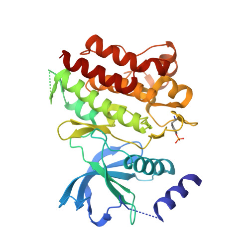
Structure and chemical inhibition of the RET tyrosine kinase domain.
Knowles, P.P., Murray-Rust, J., Kjaer, S., Scott, R.P., Hanrahan, S., Santoro, M., Ibanez, C.F., McDonald, N.Q.(2006) J Biol Chem 281: 33577-33587
- PubMed: 16928683
- DOI: https://doi.org/10.1074/jbc.M605604200
- Primary Citation of Related Structures:
2IVS, 2IVT, 2IVU, 2IVV - PubMed Abstract:
The RET proto-oncogene encodes a receptor tyrosine kinase for the glial cell line-derived neurotrophic factor family of ligands. Loss-of-function mutations in RET are implicated in Hirschsprung disease, whereas activating mutations in RET are found in human cancers, including familial medullar thyroid carcinoma and multiple endocrine neoplasias 2A and 2B. We report here the biochemical characterization of the human RET tyrosine kinase domain and the structure determination of the non-phosphorylated and phosphorylated forms. Both structures adopt the same active kinase conformation competent to bind ATP and substrate and have a pre-organized activation loop conformation that is independent of phosphorylation status. In agreement with the structural data, enzyme kinetic data show that autophosphorylation produces only a modest increase in activity. Longer forms of RET containing the juxtamembrane domain and C-terminal tail exhibited similar kinetic behavior, implying that there is no cis-inhibitory mechanism within the RET intracellular domain. Our results suggest the existence of alternative inhibitory mechanisms, possibly in trans, for the autoregulation of RET kinase activity. We also present the structures of the RET tyrosine kinase domain bound to two inhibitors, the pyrazolopyrimidine PP1 and the clinically relevant 4-anilinoquinazoline ZD6474. These structures explain why certain multiple endocrine neoplasia 2-associated RET mutants found in patients are resistant to inhibition and form the basis for design of more effective inhibitors.
Organizational Affiliation:
Structural Biology Laboratory, London Research Institute, Cancer Research UK, London WC2A 3PX, UK.