2ISC

Crystal structure of Purine Nucleoside Phosphorylase from Trichomonas vaginalis with DADMe-Imm-A


Experimental Data Snapshot

  • Method: X-RAY DIFFRACTION
  • Resolution: 2.70 Å
  • R-Value Free: 0.252 
  • R-Value Work: 0.199 
  • R-Value Observed: 0.202 

wwPDB Validation   3D Report Full Report


Ligand Structure Quality Assessment 


This is version 1.5 of the entry. See complete history


Literature

Inhibition and structure of Trichomonas vaginalis purine nucleoside phosphorylase with picomolar transition state analogues

Rinaldo-Matthis, A.Wing, C.Ghanem, M.Deng, H.Wu, P.Gupta, A.Tyler, P.C.Evans, G.B.Furneaux, R.H.Almo, S.C.Wang, C.C.Schramm, V.L.

(2007) Biochemistry 46: 659-668

  • DOI: https://doi.org/10.1021/bi061515r
  • Primary Citation of Related Structures:  
    2I4T, 2ISC

  • PubMed Abstract: 

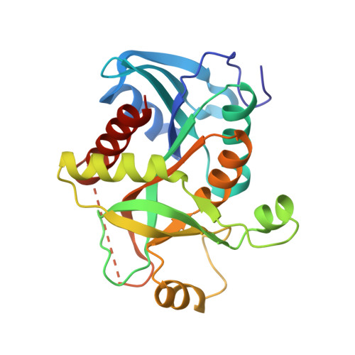
    Trichomonas vaginalis is a parasitic protozoan purine auxotroph possessing a unique purine salvage pathway consisting of a bacterial type purine nucleoside phosphorylase (PNP) and a purine nucleoside kinase. Thus, T. vaginalis PNP (TvPNP) functions in the reverse direction relative to the PNPs in other organisms. Immucillin-A (ImmA) and DADMe-Immucillin-A (DADMe-ImmA) are transition state mimics of adenosine with geometric and electrostatic features that resemble early and late transition states of adenosine at the transition state stabilized by TvPNP. ImmA demonstrates slow-onset tight-binding inhibition with TvPNP, to give an equilibrium dissociation constant of 87 pM, an inhibitor release half-time of 17.2 min, and a Km/Kd ratio of 70,100. DADMe-ImmA resembles a late ribooxacarbenium ion transition state for TvPNP to give a dissociation constant of 30 pM, an inhibitor release half-time of 64 min, and a Km/Kd ratio of 203,300. The tight binding of DADMe-ImmA supports a late SN1 transition state. Despite their tight binding to TvPNP, ImmA and DADMe-ImmA are weak inhibitors of human and P. falciparum PNPs. The crystal structures of the TvPNP x ImmA x PO4 and TvPNP x DADMe-ImmA x PO4 ternary complexes differ from previous structures with substrate analogues. The tight binding with DADMe-ImmA is in part due to a 2.7 A ionic interaction between a PO4 oxygen and the N1' cation of the hydroxypyrrolidine and is weaker in the TvPNP x ImmA x PO4 structure at 3.5 A. However, the TvPNP x ImmA x PO4 structure includes hydrogen bonds between the 2'-hydroxyl and the protein that are not present in TvPNP x DADMe-ImmA x PO4. These structures explain why DADMe-ImmA binds tighter than ImmA. Immucillin-H is a 12 nM inhibitor of TvPNP but a 56 pM inhibitor of human PNP. And this difference is explained by isotope-edited difference infrared spectroscopy with [6-18O]ImmH to establish that O6 is the keto tautomer in TvPNP x ImmH x PO4, causing an unfavorable leaving-group interaction.


  • Organizational Affiliation

    Department of Biochemistry, Albert Einstein College of Medicine, Bronx, New York 10461, USA.


Macromolecules
Find similar proteins by:  (by identity cutoff)  |  3D Structure
Entity ID: 1
MoleculeChains Sequence LengthOrganismDetailsImage
purine nucleoside phosphorylase
A, B, C, D, E
A, B, C, D, E, F
239Trichomonas vaginalisMutation(s): 0 
EC: 2.4.2.1
UniProt
Find proteins for A2E7Y6 (Trichomonas vaginalis (strain ATCC PRA-98 / G3))
Explore A2E7Y6 
Go to UniProtKB:  A2E7Y6
Entity Groups  
Sequence Clusters30% Identity50% Identity70% Identity90% Identity95% Identity100% Identity
UniProt GroupA2E7Y6
Sequence Annotations
Expand
  • Reference Sequence
Small Molecules
Ligands 2 Unique
IDChains Name / Formula / InChI Key2D Diagram3D Interactions
223
Query on 223

Download Ideal Coordinates CCD File 
I [auth A]
L [auth B]
N [auth C]
P [auth D]
R [auth E]
I [auth A],
L [auth B],
N [auth C],
P [auth D],
R [auth E],
T [auth F]
(3R,4R)-1-[(4-AMINO-5H-PYRROLO[3,2-D]PYRIMIDIN-7-YL)METHYL]-4-(HYDROXYMETHYL)PYRROLIDIN-3-OL
C12 H17 N5 O2
HYYXEHQHMXTTFP-BDAKNGLRSA-N
PO4
Query on PO4

Download Ideal Coordinates CCD File 
G [auth A]
H [auth A]
J [auth B]
K [auth B]
M [auth C]
G [auth A],
H [auth A],
J [auth B],
K [auth B],
M [auth C],
O [auth D],
Q [auth E],
S [auth F]
PHOSPHATE ION
O4 P
NBIIXXVUZAFLBC-UHFFFAOYSA-K
Binding Affinity Annotations 
IDSourceBinding Affinity
223 PDBBind:  2ISC Kd: 3.8 (nM) from 1 assay(s)
Experimental Data & Validation

Experimental Data

  • Method: X-RAY DIFFRACTION
  • Resolution: 2.70 Å
  • R-Value Free: 0.252 
  • R-Value Work: 0.199 
  • R-Value Observed: 0.202 
  • Space Group: P 21 21 21
Unit Cell:
Length ( Å )Angle ( ˚ )
a = 82.402α = 90
b = 113.821β = 90
c = 195.149γ = 90
Software Package:
Software NamePurpose
DENZOdata reduction
SCALEPACKdata scaling
REFMACrefinement
PDB_EXTRACTdata extraction
CBASSdata collection
HKL-2000data reduction
MOLREPphasing

Structure Validation

View Full Validation Report


Ligand Structure Quality Assessment 


Entry History 

Deposition Data

Revision History  (Full details and data files)

  • Version 1.0: 2007-06-05
    Type: Initial release
  • Version 1.1: 2008-05-01
    Changes: Version format compliance
  • Version 1.2: 2011-07-13
    Changes: Version format compliance
  • Version 1.3: 2017-02-01
    Changes: Structure summary
  • Version 1.4: 2017-10-18
    Changes: Refinement description
  • Version 1.5: 2024-02-21
    Changes: Data collection, Database references, Derived calculations