2IB6

Structural characterization of a blue chromoprotein and its yellow mutant from the sea anemone cnidopus japonicus


Experimental Data Snapshot

  • Method: X-RAY DIFFRACTION
  • Resolution: 2.00 Å
  • R-Value Free: 0.244 
  • R-Value Work: 0.198 
  • R-Value Observed: 0.198 

wwPDB Validation   3D Report Full Report


This is version 2.0 of the entry. See complete history


Literature

Structural Characterization of a Blue Chromoprotein and Its Yellow Mutant from the Sea Anemone Cnidopus Japonicus

Chan, M.C.Y.Karasawa, S.Mizuno, H.Bosanac, I.Ho, D.Prive, G.G.Miyawaki, A.Ikura, M.

(2006) J Biol Chem 281: 37813-37819

  • DOI: https://doi.org/10.1074/jbc.M606921200
  • Primary Citation of Related Structures:  
    2IB5, 2IB6

  • PubMed Abstract: 

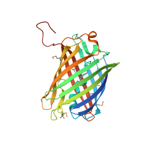
    Green fluorescent protein (GFP) and its relatives (GFP protein family) have been isolated from marine organisms such as jellyfish and corals that belong to the phylum Cnidaria (stinging aquatic invertebrates). They are intrinsically fluorescent proteins. In search of new members of the family of green fluorescent protein family, we identified a non-fluorescent chromoprotein from the Cnidopus japonicus species of sea anemone that possesses 45% sequence identity to dsRed (a red fluorescent protein). This newly identified blue color protein has an absorbance maximum of 610 nm and is hereafter referred to as cjBlue. Determination of the cjBlue 1.8 A crystal structure revealed a chromophore comprised of Gln(63)-Tyr(64)-Gly(65). The ring stacking between Tyr(64) and His(197) stabilized the cjBlue trans chromophore conformation along the Calpha2-Cbeta2 bond of 5-[(4-hydroxyphenyl)methylene]-imidazolinone, which closely resembled that of the "Kindling Fluorescent Protein" and Rtms5. Replacement of Tyr(64) with Leu in wild-type cjBlue produced a visible color change from blue to yellow with a new absorbance maximum of 417 nm. Interestingly, the crystal structure of the yellow mutant Y64L revealed two His(197) imidazole ring orientations, suggesting a flip-flop interconversion between the two conformations in solution. We conclude that the dynamics and structure of the chromophore are both essential for the optical appearance of these color proteins.


  • Organizational Affiliation

    Division of Signaling Biology, Ontario Cancer Institute and Department of Medical Biophysics, University of Toronto, Toronto, Ontario M5G 1L7, Canada.


Macromolecules
Find similar proteins by:  (by identity cutoff)  |  3D Structure
Entity ID: 1
MoleculeChains Sequence LengthOrganismDetailsImage
Yellow mutant chromo protein
A, B, C, D, E
A, B, C, D, E, F, G, H
233Epiactis japonicaMutation(s): 1 
UniProt
Find proteins for A0AQQ8 (Epiactis japonica)
Explore A0AQQ8 
Go to UniProtKB:  A0AQQ8
Entity Groups  
Sequence Clusters30% Identity50% Identity70% Identity90% Identity95% Identity100% Identity
UniProt GroupA0AQQ8
Sequence Annotations
Expand
  • Reference Sequence
Small Molecules
Ligands 1 Unique
IDChains Name / Formula / InChI Key2D Diagram3D Interactions
PO4
Query on PO4

Download Ideal Coordinates CCD File 
I [auth A]
J [auth A]
K [auth A]
L [auth B]
M [auth B]
I [auth A],
J [auth A],
K [auth A],
L [auth B],
M [auth B],
N [auth C],
O [auth D],
P [auth D],
Q [auth F],
R [auth F],
S [auth F],
T [auth G],
U [auth G],
V [auth H],
W [auth H],
X [auth H]
PHOSPHATE ION
O4 P
NBIIXXVUZAFLBC-UHFFFAOYSA-K
Modified Residues  2 Unique
IDChains TypeFormula2D DiagramParent
MSE
Query on MSE
A, B, C, D, E
A, B, C, D, E, F, G, H
L-PEPTIDE LINKINGC5 H11 N O2 SeMET
QLG
Query on QLG
A, B, C, D, E
A, B, C, D, E, F, G, H
L-PEPTIDE LINKINGC13 H18 N4 O4GLN, LEU, GLY
Experimental Data & Validation

Experimental Data

  • Method: X-RAY DIFFRACTION
  • Resolution: 2.00 Å
  • R-Value Free: 0.244 
  • R-Value Work: 0.198 
  • R-Value Observed: 0.198 
  • Space Group: P 1 21 1
Unit Cell:
Length ( Å )Angle ( ˚ )
a = 74.079α = 90
b = 126.926β = 101.97
c = 100.084γ = 90
Software Package:
Software NamePurpose
PROTEUM PLUSdata collection
CNSrefinement
PROTEUM PLUSdata reduction
PROTEUM PLUSdata scaling
CNSphasing

Structure Validation

View Full Validation Report


Entry History 

Deposition Data

Revision History  (Full details and data files)

  • Version 1.0: 2006-10-10
    Type: Initial release
  • Version 1.1: 2008-05-01
    Changes: Version format compliance
  • Version 1.2: 2011-07-13
    Changes: Derived calculations, Version format compliance
  • Version 1.3: 2023-10-25
    Changes: Data collection, Database references, Derived calculations, Refinement description
  • Version 2.0: 2023-11-15
    Changes: Advisory, Atomic model, Data collection, Derived calculations