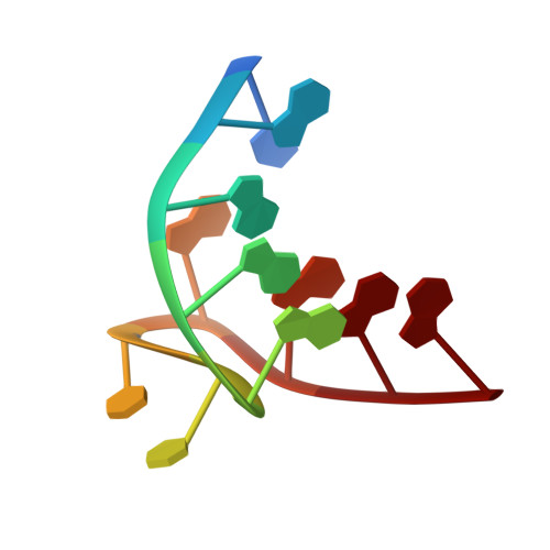
Structural basis for binding of porphyrin to human telomeres.
Parkinson, G.N., Ghosh, R., Neidle, S.(2007) Biochemistry 46: 2390-2397
- PubMed: 17274602
- DOI: https://doi.org/10.1021/bi062244n
- Primary Citation of Related Structures:
2HRI - PubMed Abstract:
Maintenance of telomere integrity is a hallmark of human cancer, and the single-stranded 3' ends of telomeric DNA are targets for small-molecule anticancer therapies. We report here the crystal structure of a bimolecular human telomeric quadruplex, of the sequence d(TAGGGTTAGGG), in a complex with the quadruplex-binding ligand 5,10,15,20-tetrakis(N-methyl-4-pyridyl)porphyrin (TMPyP4) to a resolution of 2.09 A. The DNA quadruplex topology is parallel-stranded with external double-chain-reversal propeller loops, consistent with previous structural determinations. The porphyrin molecules bind by stacking onto the TTA nucleotides, either as part of the external loop structure or at the 5' region of the stacked quadruplex. This involves stacked on hydrogen-bonded base pairs, formed from those nucleotides not involved in the formation of G-tetrads, and there are thus no direct ligand interactions with G-tetrads. This is in accord with the relative nonselectivity by TMPyP4 for quadruplex DNAs compared to duplex DNA. Porphyrin binding is achieved by remodeling of loops compared to the ligand-free structures. Implications for the design of quadruplex-binding ligands are discussed, together with a model for the formation of anaphase bridges, which are observed following cellular treatment with TMPyP4.
Organizational Affiliation:
CRUK Biomolecular Structure Group, The School of Pharmacy, University of London, 29-39 Brunswick Square, London WC1N 1AX, United Kingdom.