


Crystal Structure of P14 Alanine Variant of Antithrombin
To be published.
Experimental Data Snapshot
Entity ID: 1 | |||||
|---|---|---|---|---|---|
| Molecule | Chains | Sequence Length | Organism | Details | Image |
| Antithrombin-III | A [auth I] | 432 | Homo sapiens | Mutation(s): 2 Gene Names: SERPINC1, AT3 | 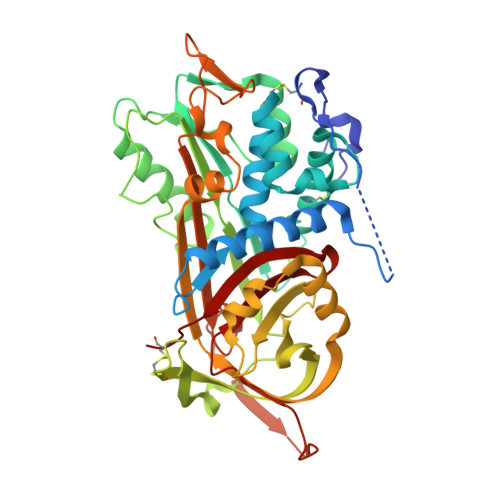 |
UniProt & NIH Common Fund Data Resources | |||||
Find proteins for P01008 (Homo sapiens) Explore P01008 Go to UniProtKB: P01008 | |||||
PHAROS: P01008 GTEx: ENSG00000117601 | |||||
Entity Groups | |||||
| Sequence Clusters | 30% Identity50% Identity70% Identity90% Identity95% Identity100% Identity | ||||
| UniProt Group | P01008 | ||||
Sequence AnnotationsExpand | |||||
| |||||
Entity ID: 2 | |||||
|---|---|---|---|---|---|
| Molecule | Chains | Sequence Length | Organism | Details | Image |
| Antithrombin-III | B [auth L] | 432 | Homo sapiens | Mutation(s): 0 | 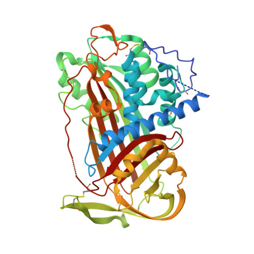 |
UniProt & NIH Common Fund Data Resources | |||||
Find proteins for P01008 (Homo sapiens) Explore P01008 Go to UniProtKB: P01008 | |||||
PHAROS: P01008 GTEx: ENSG00000117601 | |||||
Entity Groups | |||||
| Sequence Clusters | 30% Identity50% Identity70% Identity90% Identity95% Identity100% Identity | ||||
| UniProt Group | P01008 | ||||
Sequence AnnotationsExpand | |||||
| |||||
| Ligands 2 Unique | |||||
|---|---|---|---|---|---|
| ID | Chains | Name / Formula / InChI Key | 2D Diagram | 3D Interactions | |
| NAG Query on NAG | E [auth I], G [auth L], H [auth L], I [auth L] | 2-acetamido-2-deoxy-beta-D-glucopyranose C8 H15 N O6 OVRNDRQMDRJTHS-FMDGEEDCSA-N |  | ||
| GOL Query on GOL | F [auth I], J [auth L] | GLYCEROL C3 H8 O3 PEDCQBHIVMGVHV-UHFFFAOYSA-N |  | ||
| Length ( Å ) | Angle ( ˚ ) |
|---|---|
| a = 61.743 | α = 90 |
| b = 98.627 | β = 101.4 |
| c = 87.569 | γ = 90 |
| Software Name | Purpose |
|---|---|
| CNS | refinement |
| PDB_EXTRACT | data extraction |
| CNS | phasing |
RCSB PDB (citation) is hosted by
RCSB PDB is a member of the


