2H7E

Solution structure of the talin F3 domain in complex with a chimeric beta3 integrin-PIP kinase peptide- minimized average structure


Experimental Data Snapshot

  • Method: SOLUTION NMR
  • Conformers Submitted: 

wwPDB Validation   3D Report Full Report

Currently 2H7E does not have a validation slider image.


This is version 1.3 of the entry. See complete history


Literature

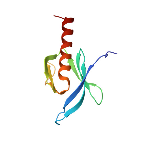
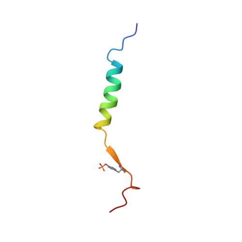
Structural basis of integrin activation by talin

Wegener, K.L.Partridge, A.W.Han, J.Pickford, A.R.Liddington, R.C.Ginsberg, M.H.Campbell, I.D.

(2007) Cell 128: 171-182

  • DOI: https://doi.org/10.1016/j.cell.2006.10.048
  • Primary Citation of Related Structures:  
    2H7D, 2H7E

  • PubMed Abstract: 

    Regulation of integrin affinity (activation) is essential for metazoan development and for many pathological processes. Binding of the talin phosphotyrosine-binding (PTB) domain to integrin beta subunit cytoplasmic domains (tails) causes activation, whereas numerous other PTB-domain-containing proteins bind integrins without activating them. Here we define the structure of a complex between talin and the membrane-proximal integrin beta3 cytoplasmic domain and identify specific contacts between talin and the integrin tail required for activation. We used structure-based mutagenesis to engineer talin and beta3 variants that interact with comparable affinity to the wild-type proteins but inhibit integrin activation by competing with endogenous talin. These results reveal the structural basis of talin's unique ability to activate integrins, identify an interaction that could aid in the design of therapeutics to block integrin activation, and enable engineering of cells with defects in the activation of multiple classes of integrins.


  • Organizational Affiliation

    Department of Biochemistry, University of Oxford, South Parks Road, Oxford OX1 3QU, England, UK.


Macromolecules
Find similar proteins by:  (by identity cutoff)  |  3D Structure
Entity ID: 1
MoleculeChains Sequence LengthOrganismDetailsImage
Talin-1101Gallus gallusMutation(s): 1 
Gene Names: TLN1TLN
UniProt
Find proteins for P54939 (Gallus gallus)
Explore P54939 
Go to UniProtKB:  P54939
Entity Groups  
Sequence Clusters30% Identity50% Identity70% Identity90% Identity95% Identity100% Identity
UniProt GroupP54939
Sequence Annotations
Expand
  • Reference Sequence
Find similar proteins by:  (by identity cutoff)  |  3D Structure
Entity ID: 2
MoleculeChains Sequence LengthOrganismDetailsImage
Chimera of 24-mer peptide from Integrin beta-3 and 10-mer peptide from Phosphatidylinositol-4-phosphate 5-kinase type-1 gamma34N/AMutation(s): 1 
UniProt & NIH Common Fund Data Resources
Find proteins for O70161 (Mus musculus)
Explore O70161 
Go to UniProtKB:  O70161
IMPC:  MGI:1298224
Entity Groups  
Sequence Clusters30% Identity50% Identity70% Identity90% Identity95% Identity100% Identity
UniProt GroupO70161
Sequence Annotations
Expand
  • Reference Sequence
Small Molecules
Modified Residues  1 Unique
IDChains TypeFormula2D DiagramParent
PTR
Query on PTR
B
L-PEPTIDE LINKINGC9 H12 N O6 PTYR
Experimental Data & Validation

Experimental Data

  • Method: SOLUTION NMR
  • Conformers Submitted: 

Structure Validation

View Full Validation Report

Currently 2H7E does not have a validation slider image.


Entry History 

Deposition Data

Revision History  (Full details and data files)

  • Version 1.0: 2007-01-30
    Type: Initial release
  • Version 1.1: 2008-05-01
    Changes: Version format compliance
  • Version 1.2: 2011-07-13
    Changes: Version format compliance
  • Version 1.3: 2021-11-10
    Changes: Database references, Derived calculations