Novel Chromophores and Buried Charges Control Color in mFruits
Shu, X., Shaner, N.C., Yarbrough, C.A., Tsien, R.Y., Remington, S.J.(2006) Biochemistry 45: 9639-9647
- PubMed: 16893165
- DOI: https://doi.org/10.1021/bi060773l
- Primary Citation of Related Structures:
2H5O, 2H5P, 2H5Q, 2H5R, 2H8Q - PubMed Abstract:
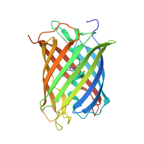
mFruits are second-generation monomeric red fluorescent proteins (mRFPs) that have improved brightness and photostability compared to the first-generation mRFP1. The emission and excitation maxima are distributed over the remarkably large ranges of about 550-650 and 540-590 nm, respectively; however, the variations in the spectra can be traced to a few key amino acids. Spectroscopic and atomic resolution crystallographic analyses of three representatives, mOrange, mStrawberry, and mCherry, reveal that different mechanisms operate to establish the excitation and emission maxima. Evidently, they all undergo the second oxidation step to produce an acylimine linkage in the polypeptide backbone. In comparison to the progenitor DsRed, direct covalent modification to this linkage (mOrange) and indirect modification of the chromophore environment (mStrawberry and mCherry) produce strong blue- and red-shifted variants. The blue shift of mOrange is induced by an unprecedented covalent modification of the protein backbone. The electron-density map indicates the formation of a third heterocycle, 2-hydroxy-dihydrooxazole, upon the reaction of Thr 66 Ogamma with the polypeptide backbone, which in turn reduces the conjugation of the carbonyl at position 65 with the rest of the chromophore. In mStrawberry and mCherry, the movement of charged Lys 70 and protonation of Glu 215 are proposed to modify the chromophore electron-density distribution, inducing the red shift. pH-dependent spectral shifts of mCherry and mStrawberry appear to result from the titration of Glu 215, although, for mStrawberry, partial cyclization of Thr 66 may contribute at high pH.
Organizational Affiliation:
Institute of Molecular Biology and Department of Physics, University of Oregon, Eugene, Oregon 97403, USA.