2H1P

THE THREE-DIMENSIONAL STRUCTURES OF A POLYSACCHARIDE BINDING ANTIBODY TO CRYPTOCOCCUS NEOFORMANS AND ITS COMPLEX WITH A PEPTIDE FROM A PHAGE DISPLAY LIBRARY: IMPLICATIONS FOR THE IDENTIFICATION OF PEPTIDE MIMOTOPES


Experimental Data Snapshot

  • Method: X-RAY DIFFRACTION
  • Resolution: 2.40 Å
  • R-Value Work: 0.186 

wwPDB Validation   3D Report Full Report


This is version 1.3 of the entry. See complete history


Literature

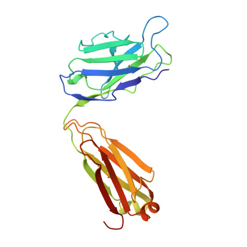
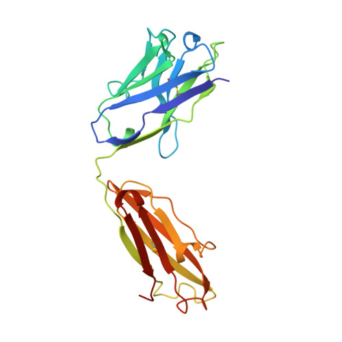
The three-dimensional structures of a polysaccharide binding antibody to Cryptococcus neoformans and its complex with a peptide from a phage display library: implications for the identification of peptide mimotopes.

Young, A.C.Valadon, P.Casadevall, A.Scharff, M.D.Sacchettini, J.C.

(1997) J Mol Biol 274: 622-634

  • DOI: https://doi.org/10.1006/jmbi.1997.1407
  • Primary Citation of Related Structures:  
    2H1P

  • PubMed Abstract: 

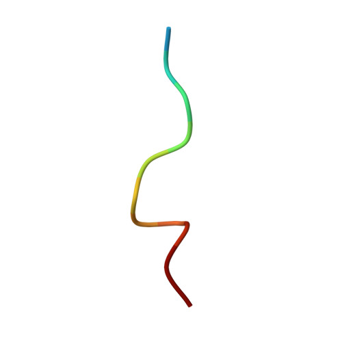
    The three-dimensional structure of 2H1, a protective monoclonal antibody to Cryptococcus neoformans, has been solved at 2.4 A resolution, in both its unbound form and in complex with the 12 amino acid residue peptide PA1 (GLQYTPSWMLVG). PA1 was previously identified as a potential mimotope of the cryptococcal capsular polysaccharide by screening of a phage display peptide library. Peptide binding is associated with only minor rearrangements of some side-chains and a small shift in the H2 loop of the antibody. The peptide assumes a tightly coiled conformation consisting of one inverse gamma-turn and one type II beta-turn that serves to place the entire peptide motif, consisting of ThrP5, ProP6, TrpP8, MetP9 and LeuP10, into a depression in the antibody combining site. A small number of H-bonds between peptide and antibody contribute to the affinity and specificity. Poor steric complementarity between PA1 and the antibody heavy chain along with the fact that the majority of the interactions between 2H1 and PA1 involve van der Waals interactions with the light chain may explain why this peptide acts as only a partial mimotope of the capsular polysaccharide epitope.


  • Organizational Affiliation

    Department of Microbiology, Albert Einstein College of Medicine, Bronx, NY 10461, USA.


Macromolecules
Find similar proteins by:  (by identity cutoff)  |  3D Structure
Entity ID: 1
MoleculeChains Sequence LengthOrganismDetailsImage
2H1A [auth L]219Mus musculusMutation(s): 0 
Entity Groups  
Sequence Clusters30% Identity50% Identity70% Identity90% Identity95% Identity100% Identity
Sequence Annotations
Expand
  • Reference Sequence
Find similar proteins by:  (by identity cutoff)  |  3D Structure
Entity ID: 2
MoleculeChains Sequence LengthOrganismDetailsImage
2H1B [auth H]220Mus musculusMutation(s): 0 
Entity Groups  
Sequence Clusters30% Identity50% Identity70% Identity90% Identity95% Identity100% Identity
Sequence Annotations
Expand
  • Reference Sequence

Find similar proteins by:  Sequence   |   3D Structure  

Entity ID: 3
MoleculeChains Sequence LengthOrganismDetailsImage
PA1C [auth P]12N/AMutation(s): 0 
Entity Groups  
Sequence Clusters30% Identity50% Identity70% Identity90% Identity95% Identity100% Identity
Sequence Annotations
Expand
  • Reference Sequence
Experimental Data & Validation

Experimental Data

  • Method: X-RAY DIFFRACTION
  • Resolution: 2.40 Å
  • R-Value Work: 0.186 
  • Space Group: P 1
Unit Cell:
Length ( Å )Angle ( ˚ )
a = 43.79α = 91.82
b = 51.25β = 98.57
c = 60.18γ = 107.84
Software Package:
Software NamePurpose
SAINTdata scaling
SAINTdata reduction
X-PLORmodel building
TNTrefinement
X-PLORrefinement
X-PLORphasing

Structure Validation

View Full Validation Report


Entry History 

Deposition Data

Revision History  (Full details and data files)

  • Version 1.0: 1998-01-28
    Type: Initial release
  • Version 1.1: 2008-03-24
    Changes: Version format compliance
  • Version 1.2: 2011-07-13
    Changes: Version format compliance
  • Version 1.3: 2023-08-09
    Changes: Database references, Refinement description