2GZH

Crystal Structure of Rab11 in Complex with Rab11-Family Interacting Protein 2


Experimental Data Snapshot

  • Method: X-RAY DIFFRACTION
  • Resolution: 2.47 Å
  • R-Value Free: 0.254 
  • R-Value Work: 0.197 
  • R-Value Observed: 0.199 

wwPDB Validation   3D Report Full Report


This is version 1.3 of the entry. See complete history


Literature

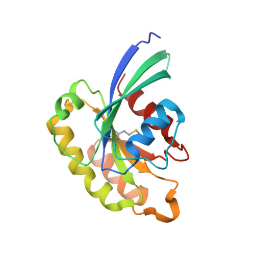
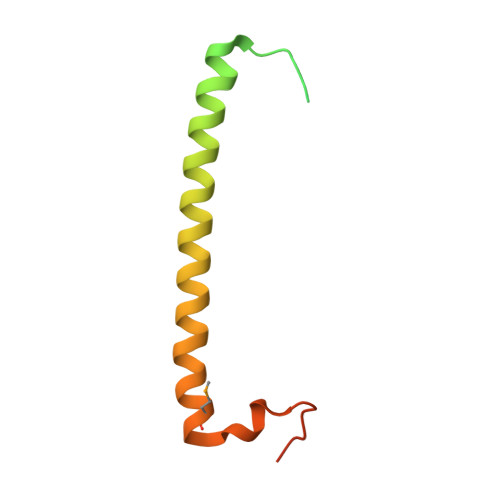
Crystal structure of rab11 in complex with rab11 family interacting protein 2.

Jagoe, W.N.Lindsay, A.J.Read, R.J.McCoy, A.J.McCaffrey, M.W.Khan, A.R.

(2006) Structure 14: 1273-1283

  • DOI: https://doi.org/10.1016/j.str.2006.06.010
  • Primary Citation of Related Structures:  
    2GZD, 2GZH

  • PubMed Abstract: 

    The small GTPase Rab11 regulates the recycling of endosomes to the plasma membrane via interactions with the Rab11 family of interacting proteins (FIPs). FIPs contain a highly conserved Rab binding domain (RBD) at their C termini whose structure is unknown. Here, we have determined the crystal structure of the RBD of FIP2 in complex with Rab11(GTP) by single wavelength anomalous diffraction methods. The overall structure is a heterotetramer with dyad symmetry, arranged as a Rab11-(FIP2)2-Rab11 complex. FIP2 forms a central alpha-helical coiled coil, with both helices contributing to the Rab11 binding patch on equivalent and opposite sides of the homodimer. Switch 1 of Rab11 is embedded between the two helices, while switch 2 remains flexible and is peripherally associated with the effector. The complex reveals the structural basis for Rab11 recognition by FIPs and suggests the molecular mechanisms underlying endocytic recycling pathways.


  • Organizational Affiliation

    School of Biochemistry and Immunology, Trinity College, Dublin 2, Ireland.


Macromolecules
Find similar proteins by:  (by identity cutoff)  |  3D Structure
Entity ID: 1
MoleculeChains Sequence LengthOrganismDetailsImage
Ras-related protein Rab-11A173Homo sapiensMutation(s): 2 
Gene Names: RAB11a
EC: 3.6.5.2
UniProt & NIH Common Fund Data Resources
Find proteins for P62491 (Homo sapiens)
Explore P62491 
Go to UniProtKB:  P62491
PHAROS:  P62491
GTEx:  ENSG00000103769 
Entity Groups  
Sequence Clusters30% Identity50% Identity70% Identity90% Identity95% Identity100% Identity
UniProt GroupP62491
Sequence Annotations
Expand
  • Reference Sequence
Find similar proteins by:  (by identity cutoff)  |  3D Structure
Entity ID: 2
MoleculeChains Sequence LengthOrganismDetailsImage
RAB11-FIP2 long isoform107Homo sapiensMutation(s): 1 
UniProt & NIH Common Fund Data Resources
Find proteins for Q7L804 (Homo sapiens)
Explore Q7L804 
Go to UniProtKB:  Q7L804
PHAROS:  Q7L804
GTEx:  ENSG00000107560 
Entity Groups  
Sequence Clusters30% Identity50% Identity70% Identity90% Identity95% Identity100% Identity
UniProt GroupQ7L804
Sequence Annotations
Expand
  • Reference Sequence
Small Molecules
Experimental Data & Validation

Experimental Data

  • Method: X-RAY DIFFRACTION
  • Resolution: 2.47 Å
  • R-Value Free: 0.254 
  • R-Value Work: 0.197 
  • R-Value Observed: 0.199 
  • Space Group: P 31 2 1
Unit Cell:
Length ( Å )Angle ( ˚ )
a = 64.74α = 90
b = 64.74β = 90
c = 112.4γ = 120
Software Package:
Software NamePurpose
REFMACrefinement
ADSCdata collection
HKL-2000data scaling
PHASERphasing

Structure Validation

View Full Validation Report


Entry History 

Deposition Data

  • Released Date: 2006-08-15 
  • Deposition Author(s): Khan, A.R.

Revision History  (Full details and data files)

  • Version 1.0: 2006-08-15
    Type: Initial release
  • Version 1.1: 2008-05-01
    Changes: Version format compliance
  • Version 1.2: 2011-07-13
    Changes: Version format compliance
  • Version 1.3: 2021-10-20
    Changes: Database references, Derived calculations