2GW3

Crystal structure of stony coral fluorescent protein Kaede, green form


Experimental Data Snapshot

  • Method: X-RAY DIFFRACTION
  • Resolution: 1.40 Å
  • R-Value Free: 0.214 
  • R-Value Work: 0.197 

wwPDB Validation   3D Report Full Report


This is version 2.0 of the entry. See complete history


Literature

Crystallographic evidence for water-assisted photo-induced peptide cleavage in the stony coral fluorescent protein Kaede.

Hayashi, I.Mizuno, H.Tong, K.I.Furuta, T.Tanaka, F.Yoshimura, M.Miyawaki, A.Ikura, M.

(2007) J Mol Biol 372: 918-926

  • DOI: https://doi.org/10.1016/j.jmb.2007.06.037
  • Primary Citation of Related Structures:  
    2GW3, 2GW4

  • PubMed Abstract: 

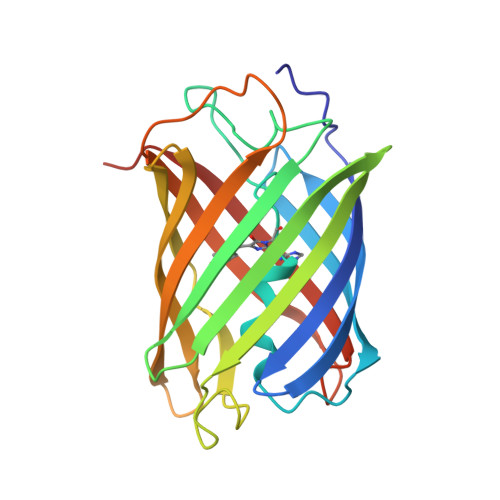
    A coral fluorescent protein from Trachyphyllia geoffroyi, Kaede, possesses a tripeptide of His62-Tyr63-Gly64, which forms a chromophore with green fluorescence. This chromophore's fluorescence turns red following UV light irradiation. We have previously shown that such photoconversion is achieved by a formal beta-elimination reaction, which results in a cleavage of the peptide bond found between the amide nitrogen and the alpha-carbon at His62. However, the stereochemical arrangement of the chromophore and the precise structural basis for this reaction mechanism previously remained unknown. Here, we report the crystal structures of the green and red form of Kaede at 1.4 A and 1.6 A resolutions, respectively. Our structures depict the cleaved peptide bond in the red form. The chromophore conformations both in the green and red forms are similar, except a well-defined water molecule in the proximity of the His62 imidazole ring in the green form. We propose a molecular mechanism for green-to-red photoconversion, which is assisted by the water molecule.


  • Organizational Affiliation

    Division of Signaling Biology, Ontario Cancer Institute and Department of Medical Biophysics, 101 College St., Toronto, Ontario, M5G 1L7, Canada.


Macromolecules
Find similar proteins by:  (by identity cutoff)  |  3D Structure
Entity ID: 1
MoleculeChains Sequence LengthOrganismDetailsImage
Kaede
A, B
225Trachyphyllia geoffroyiMutation(s): 1 
UniProt
Find proteins for Q8I6J8 (Trachyphyllia geoffroyi)
Explore Q8I6J8 
Go to UniProtKB:  Q8I6J8
Entity Groups  
Sequence Clusters30% Identity50% Identity70% Identity90% Identity95% Identity100% Identity
UniProt GroupQ8I6J8
Sequence Annotations
Expand
  • Reference Sequence
Experimental Data & Validation

Experimental Data

  • Method: X-RAY DIFFRACTION
  • Resolution: 1.40 Å
  • R-Value Free: 0.214 
  • R-Value Work: 0.197 
  • Space Group: C 1 2 1
Unit Cell:
Length ( Å )Angle ( ˚ )
a = 132.496α = 90
b = 80.476β = 113.82
c = 53.528γ = 90
Software Package:
Software NamePurpose
ADSCdata collection
HKL-2000data reduction
CNSrefinement
HKL-2000data scaling
CNSphasing

Structure Validation

View Full Validation Report


Entry History 

Deposition Data

Revision History  (Full details and data files)

  • Version 1.0: 2007-05-08
    Type: Initial release
  • Version 1.1: 2007-11-14
    Changes: Version format compliance
  • Version 1.2: 2011-07-13
    Changes: Derived calculations, Version format compliance
  • Version 2.0: 2023-11-15
    Changes: Atomic model, Data collection, Database references, Derived calculations