2GSY

The 2.6A structure of Infectious Bursal Virus Derived T=1 Particles


Experimental Data Snapshot

  • Method: X-RAY DIFFRACTION
  • Resolution: 2.60 Å
  • R-Value Free: 0.267 
  • R-Value Work: 0.254 
  • R-Value Observed: 0.255 

wwPDB Validation   3D Report Full Report


This is version 1.3 of the entry. See complete history


Literature

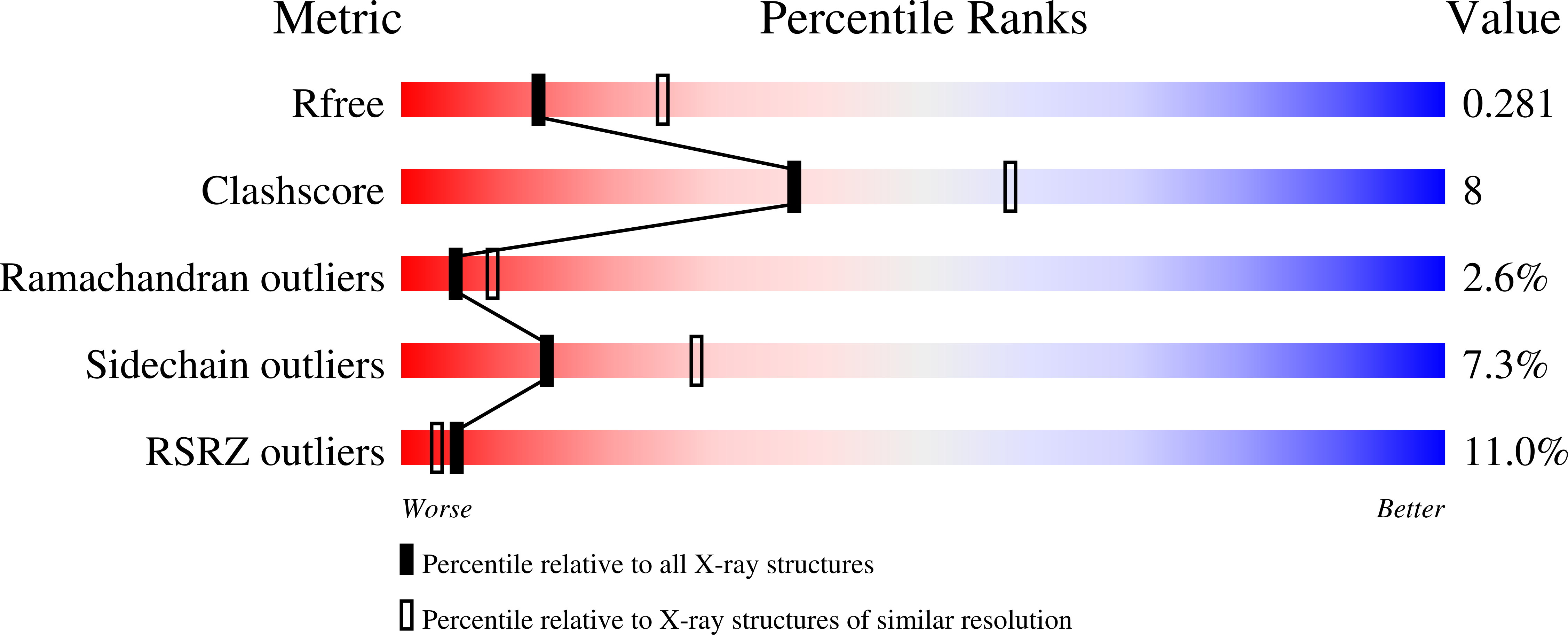
The 2.6-angstrom structure of infectious bursal disease virus-derived t=1 particles reveals new stabilizing elements of the virus capsid.

Garriga, D.Querol-Audi, J.Abaitua, F.Saugar, I.Pous, J.Verdaguer, N.Caston, J.R.Rodriguez, J.F.

(2006) J Virol 80: 6895-6905

  • DOI: https://doi.org/10.1128/JVI.00368-06
  • Primary Citation of Related Structures:  
    2GSY

  • PubMed Abstract: 

    Infectious bursal disease virus (IBDV), a member of the Birnaviridae family, is a double-stranded RNA virus that causes a highly contagious disease in young chickens leading to significant economic losses in the poultry industry. The VP2 protein, the only structural component of the IBDV icosahedral capsid, spontaneously assembles into T=1 subviral particles (SVP) when individually expressed as a chimeric gene. We have determined the crystal structure of the T=1 SVP to 2.60 A resolution. Our results show that the 20 trimeric VP2 clusters forming the T=1 shell are further stabilized by calcium ions located at the threefold icosahedral axes. The structure also reveals a new unexpected domain swapping that mediates interactions between adjacent trimers: a short helical segment located close to the end of the long C-terminal arm of VP2 is projected toward the threefold axis of a neighboring VP2 trimer, leading to a complex network of interactions that increases the stability of the T=1 particles. Analysis of crystal packing shows that the exposed capsid residues, His253 and Thr284, determinants of IBDV virulence and the adaptation of the virus to grow in cell culture, are involved in particle-particle interactions.


  • Organizational Affiliation

    Institut de Biologia Molecular Barcelona, CSIC, Josep Samitier 1-5, 08028 Barcelona, Spain.


Macromolecules
Find similar proteins by:  (by identity cutoff)  |  3D Structure
Entity ID: 1
MoleculeChains Sequence LengthOrganismDetailsImage
polyprotein
A, B, C, D, E
A, B, C, D, E, F, G, H, I, J, K, L, M, N, O, P, Q, R, S, T
456Infectious bursal disease virusMutation(s): 0 
UniProt
Find proteins for P61825 (Avian infectious bursal disease virus)
Explore P61825 
Go to UniProtKB:  P61825
Entity Groups  
Sequence Clusters30% Identity50% Identity70% Identity90% Identity95% Identity100% Identity
UniProt GroupP61825
Sequence Annotations
Expand
  • Reference Sequence
Small Molecules
Ligands 1 Unique
IDChains Name / Formula / InChI Key2D Diagram3D Interactions
CA
Query on CA

Download Ideal Coordinates CCD File 
AA [auth L]
BA [auth M]
CA [auth O]
DA [auth S]
EA [auth T]
AA [auth L],
BA [auth M],
CA [auth O],
DA [auth S],
EA [auth T],
U [auth A],
V [auth B],
W [auth C],
X [auth D],
Y [auth F],
Z [auth G]
CALCIUM ION
Ca
BHPQYMZQTOCNFJ-UHFFFAOYSA-N
Experimental Data & Validation

Experimental Data

  • Method: X-RAY DIFFRACTION
  • Resolution: 2.60 Å
  • R-Value Free: 0.267 
  • R-Value Work: 0.254 
  • R-Value Observed: 0.255 
  • Space Group: P 21 3
Unit Cell:
Length ( Å )Angle ( ˚ )
a = 326.3α = 90
b = 326.3β = 90
c = 326.3γ = 90
Software Package:
Software NamePurpose
REFMACrefinement
MOSFLMdata reduction
AMoREphasing

Structure Validation

View Full Validation Report


Entry History 

Deposition Data

Revision History  (Full details and data files)

  • Version 1.0: 2006-07-18
    Type: Initial release
  • Version 1.1: 2008-05-01
    Changes: Version format compliance
  • Version 1.2: 2011-07-13
    Changes: Derived calculations, Version format compliance
  • Version 1.3: 2023-08-30
    Changes: Data collection, Database references, Derived calculations, Refinement description